Straumann® Ceramic Healing Abutments

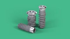
The first step to harmonious soft-tissue healing.

The Straumann® Ceramic Healing Abutments offer favorable conditions for soft-tissue attachment, hereby supporting a healthy peri-implant environment. Their well-proven zirconia material also helps surgeons and prosthodontists looking for less plaque attachment and enhanced soft tissue healing from the day of surgery.

Key indications
Single-unit
Fixation
Material
ZrO2
Workflow
Traditional
Implant Systems
BL | BLT
Implant Connections
NC, RC
Favorable soft-tissue attachment

In general, more favorable soft tissue attachment around zirconia than around titanium, with blood circulation similar to that around natural tooth.1-2

Designed for healthy peri-implant environment

Less plaque attachment on zirconia due to smoother surface compared to titanium.2-3,8-9 

Ease of use

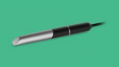
Aspiration-security thanks to integrated screw.
Color-coding to clearly identify the corresponding prosthetic platform.

Esthetics from the day of surgery

Ceramic abutments for the healing phase. 
Final restoration using Straumann® Cares® ceramic options.

Brochures and videos

Looking for additional information? You'll find them in the Download Center.

Featured stories on youTooth

Contact us

Data usage agreement*

Read our privacy notice.

Please retype the code above

Downloads

Documents

Videos & Animations

Scientific evidence

Catalogs

References

1 Kajiwara N, Masaki C, Mukaibo T, Kondo Y, Nakamoto T, Hosokawa R (2015). Soft tissue biological response to zirconia and metal implant abutments compared with natural tooth: microcirculation monitoring as a novel bioindicator. Implant Dent 24(1):37-41. 2 Degidi M, Artese L, Scarano A, Perrotti V, Gehrke P, Piattelli A. Inflammatory infiltrate, microvessel density, nitric oxide synthase expression, vascular endothelial growth factor expression, and proliferative activity in peri-implant soft tissues around titanium and zirconium oxide healing caps. J Periodontol. 2006 Jan;77(1):73-80. 3 Liñares A, Grize L, Muñoz F, Pippenger BE, Dard M, Domken O, Blanco-Carrión J. Histological assessment of hard and soft tissues surrounding a novel ceramic implant: a pilot study in the minipig. J Clin Periodontol. 2016 Jun;43(6):538-46.  4 Erbshäuser M., Zirconium dioxide dental implants in single-tooth gaps – An alternative to titanium? (Article in German). Implantologie Journal 11 | 2015, 32-36. 5 Welander M, Abrahamsson I, Berglundh T. The mucosal barrier at implant abutments of different materials. Clin Oral Implants Res. 2008 Jul;19(7):635-41. 6 Tetè S, Mastrangelo F, Bianchi A, Zizzari V, Scarano A. Collagen fiber orientation around machined titanium and zirconia dental implant necks: an animal study. Int J Oral Maxillofac Implants. 2009 Jan-Feb;24(1):52-8. 7 De Medeiros RA, Vechiato-Filho AJ, Pellizzer EP, Mazaro JV, dos Santos DM, Goiato MC (2013). Analysis of the peri-implant soft tissues in contact with zirconia abutments: an evidence-based literature review. J Contemp Dent Pract 14(3):567- 572. 8 S. Roehling, M. Astasov-Frauenhoffer, I. Hauser-Gerspach, O. Braissant, H. Engelhardt, T. Waltimo, M. Gahlert. In vitro biofilm formation on commercially available machined and micro-roughened titanium and zirconia implant surfaces. Clin. Oral Impl. Res. 26 (Suppl. 12), 2015. 9 Scarano A, Piattelli M, Caputi S, Favero GA, Piattelli A. Bacterial adhesion on commercially pure titanium and zirconium oxide disks: an in vivo human study. J Periodontol. 2004 Feb;75(2):292-6.