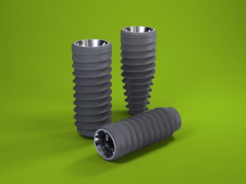
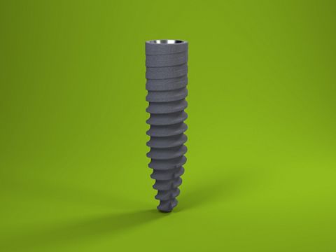
Straumann® Bone Level Tapered Ø 2.9 mm implant in the esthetic zone: A 16-month follow-up
A clinical case report by Artur Caleres, Portugal
This case report describes treatment with a Straumann® Bone Level Tapered (BLT) Ø 2.9 mm implant for agenesis of a maxillary lateral incisor. The patient was opposed to any form of orthodontic treatment. This clinical case would have been difficult to manage without this kind of implant. It can be concluded that Straumann® BLT Ø 2.9 mm implants represent a safe and reliable solution for narrow interdental gaps.
Initial situation
A healthy 26-year-old female patient with lateral incisor agenesis wanted a fixed solution for the missing tooth. She had good oral hygiene and was wearing an acrylic Maryland bridge as a provisional restoration. (Fig. 1-2)
Regarding her smoking status, she was considered to be a light smoker (three to four cigarettes a week).
According to the patient, she underwent a 4-year orthodontic treatment. Therefore, she wanted treatment without any kind of orthodontics, nor any compromise of neighboring teeth.
After an esthetic evaluation, it was concluded that she had a medium smile line (Fig. 3), rotation of tooth 13, an overjet of 4 mm, and generalized gingival recession that was more pronounced in the canine area. Treating this gingival recession was not an immediate priority, although it was in the patient’s future interests.
Treatment planning
After an assessment of periapical radiography (Fig. 6), cone beam computed tomography (Fig. 7-8) and a clinical evaluation of tooth 12, it was decided that a Straumann® BLT Ø 2.9 mm was a good solution for this clinical case due to the narrow interdental space and limited horizontal bone available (5.25 mm).
Although there was enough bone volume for implant placement, there was not enough soft tissue. (Fig. 9-10)
For this reason, it was decided to use a connective tissue graft from the tuberosity. Recent studies have demonstrated that tissue from the tuberosity is very dense and coarse and seems to contain more collagen and less fat and glandular tissue than does tissue from the palatal area. Additionally, healing is usually less painful for the patient (Zuhr O 2014).
Surgical procedure
After a crestal incision and a vertical incision distal to tooth 13 (Fig. 11), a flap was raised. The surgical template (Fig. 12) was based on a wax-up. After using the recommended drill sequence, a Straumann® BLT Ø 2.9 mm was placed at the right lateral incisor position with an insertion torque of 20 Ncm. After this, a closure cap was placed. (Fig. 13-14).
A subepithelial connective tissue graft was collected from the tuberosity and stabilized on the recipient area using a 5.0 PGA absorbable suture. (Fig. 15) A 5.0 polyamide suture was then used to close the wound. (Fig. 16-17)
A provisional acrylic Maryland tooth was bonded to the adjacent teeth for the subsequent healing period (4 months) (Fig. 18) and a final periapical radiograph was taken after surgery using a Kodak RVG 5200 sensor (Carestream®). (Fig. 19)
The first week of the healing period passed without any complications. (Fig. 20-21)
Prosthetic procedure
Some 4 months after surgery, a minimal access incision to the implant was made. (Fig. 22)
A screw-retained provisional crown was screwed to the implant via a provisional titanium abutment with a 3 mm transmucosal height. (Fig. 23-24)
This provisional solution was left in place for 3 months. (Fig. 25-26)
Lastly, a polyvinyl siloxane impression was made of the provisional crown profile. (Fig. 27-28)
A single partially layered ceramic/zirconia crown was cemented in place 2 weeks later via a titanium abutment with a 2 mm transmucosal height. (Fig. 29-30)
The connective tissue graft from the tuberosity led to a stable result with an increase in soft tissue volume. (Fig. 31-32)
The final images show the clinical situation at 16 months of follow-up after implant insertion. A periapical radiograph was taken using a Kodak RVG 5200 sensor (Carestream®) during this visit, in which it is possible to see the stability of the tissues. (Fig. 33-34)
Treatment Outcome
Here we used a Straumann® BLT Ø 2.9 mm implant in a narrow interdental gap and unfavorable interradicular space combined with mucogingival surgery and the placement of a provisional crown before the insertion of the final crown. The implant resulted in a successful outcome, improving soft tissue stability and esthetics.


