Fully edentulous treatment with digital planning and guided surgery to achieve the optimum restoratively driven outcome
P. Garrett, K. Trobough, R. Sugita and A. Pitz, Texas/USA
Initial situation
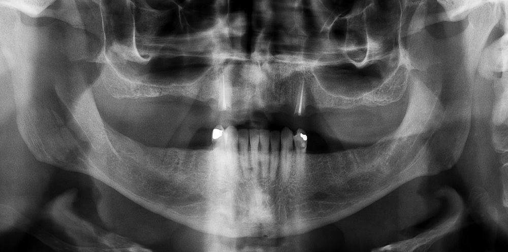
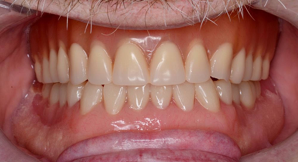
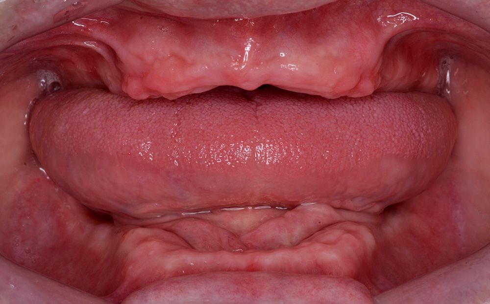
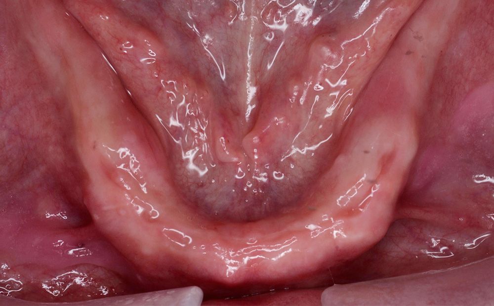
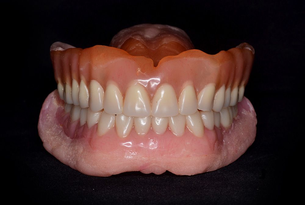
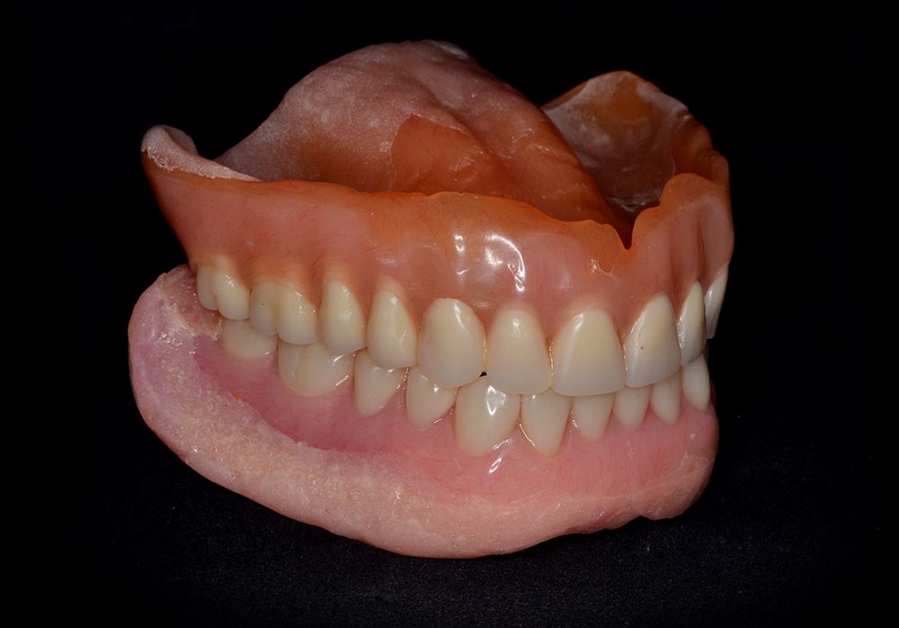
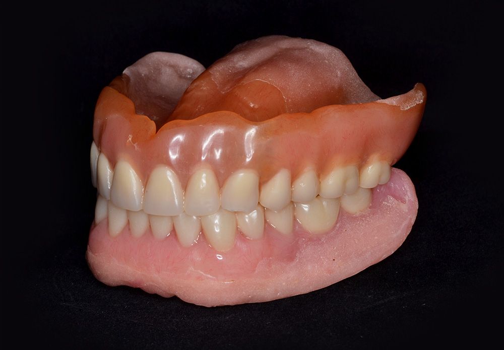
A male patient, aged 59, presented with a previously fabricated maxillary overdenture and failing maxillary and mandibular dentition (Fig. 1). The patient requested a conventional removable prosthesis for the maxillary arch and a fixed option to replace his mandibular dentition. His previous maxillary overdenture had poor retention and was planned to be remade. Full-arch extractions were completed three months prior to implant placement. An immediate denture was fabricated and delivered on the day of the extractions.
Treatment plan
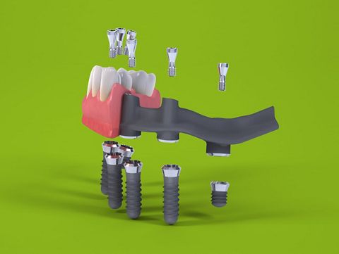
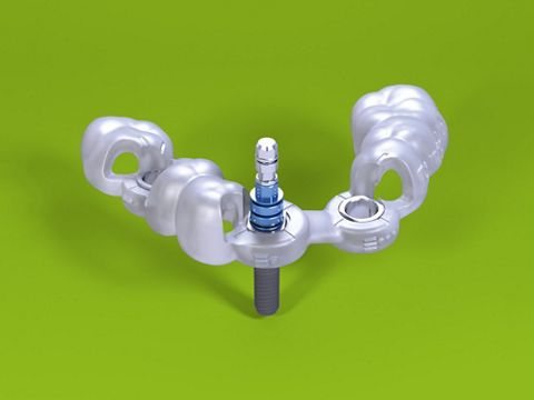
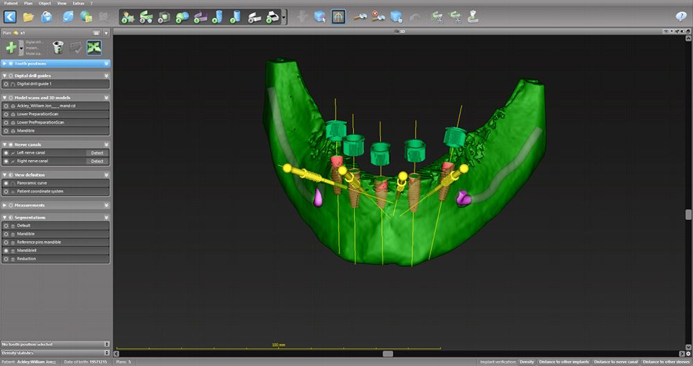
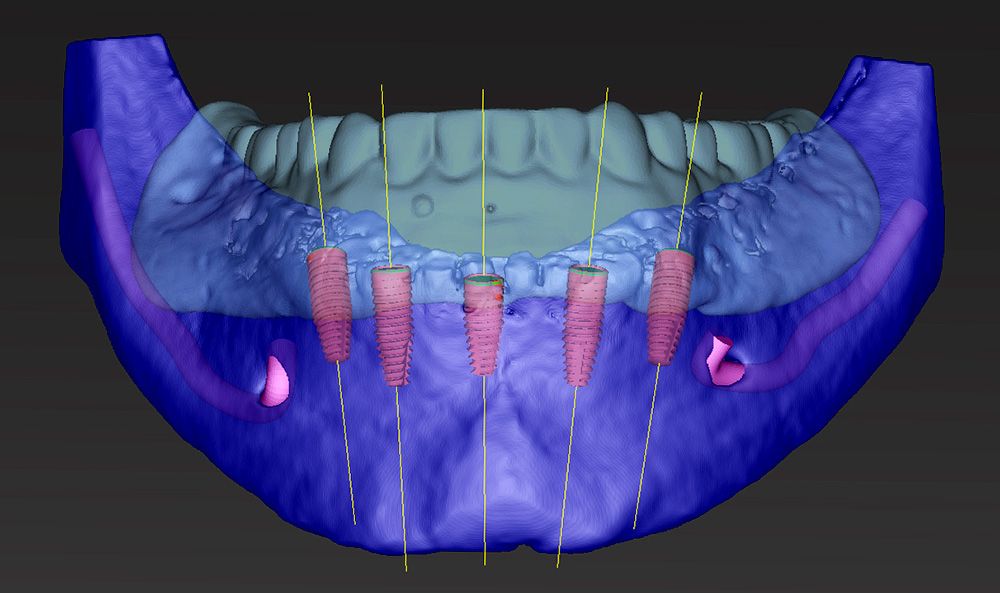
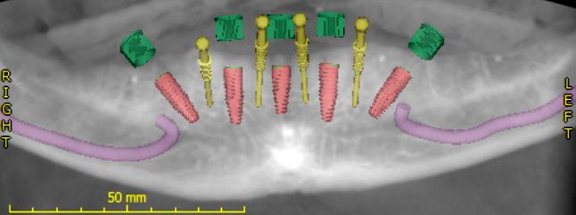
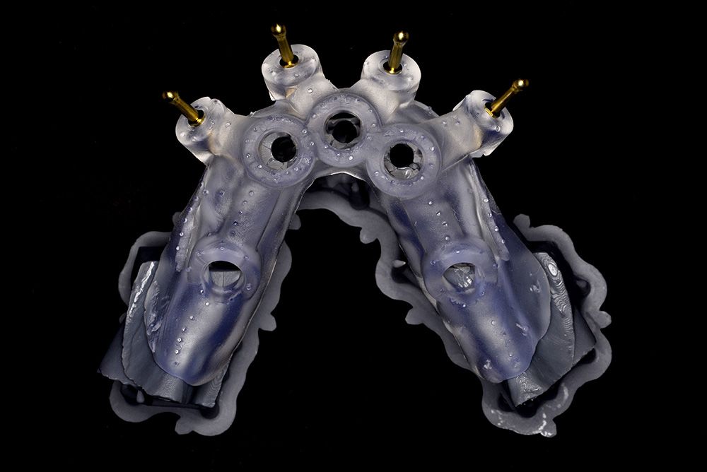
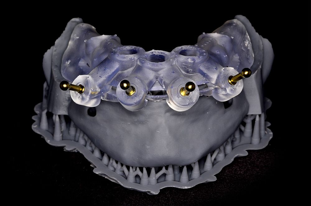
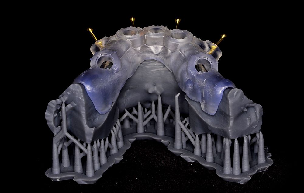
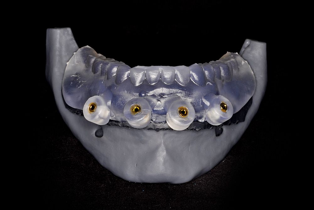
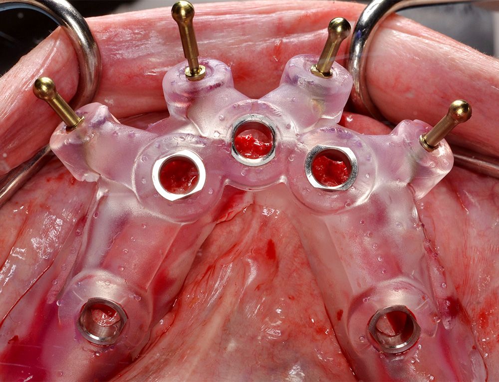
The treatment plan involved the placement of five Straumann® mandibular implants to support a fixed hybrid prosthesis. Four of the dental implants were to be used for the provisional fixed prosthesis to attain cross-arch stabilization during osseointegration. The patient was referred for a CT scan using a dual scan protocol with coDiagnostiX™ (Figs. 2-4). The virtual planning strategy was to bypass the mandibular canals and mental foramina and make use of all available bone by using a predictable procedure that remained simple and affordable for the patient. Once designed, the guide and prosthesis STL files were imported into a separate CAD program to design the creation of the “occlusion fixation guide”. Both the surgical guide (Figs. 5-7) and the occlusion fixation guide (Fig. 8) were fabricated using additive manufacturing techniques.
Surgical procedure
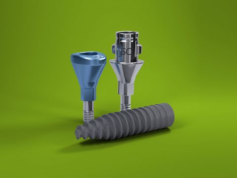
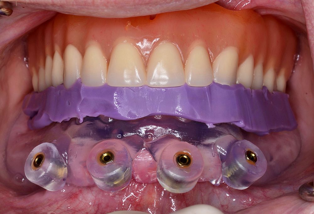
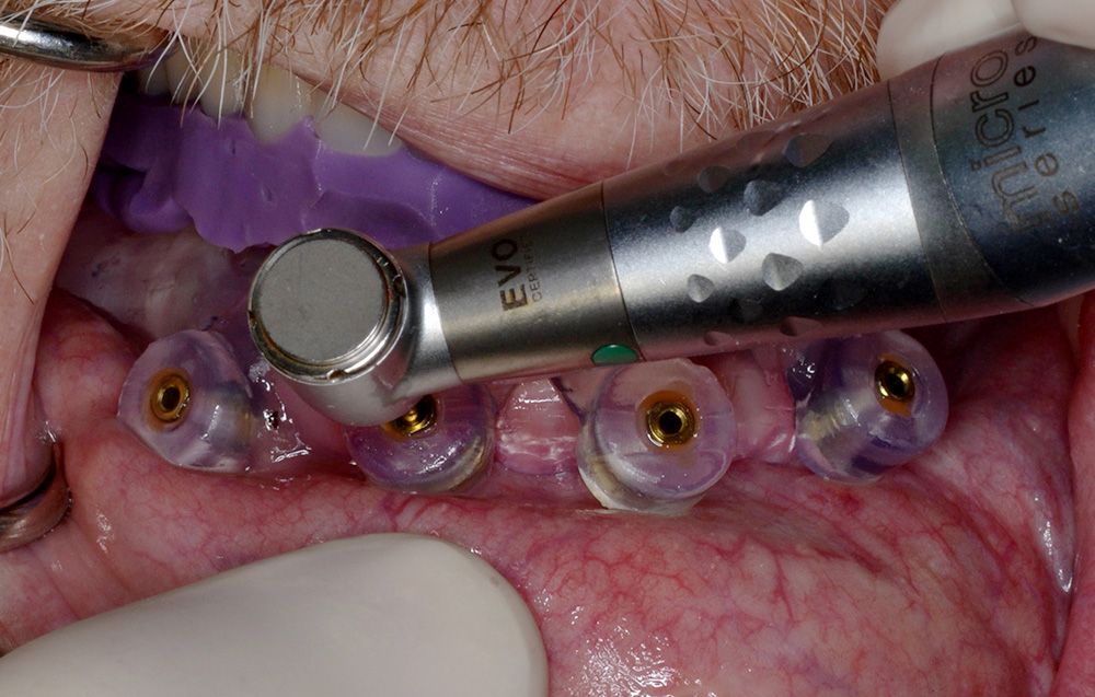
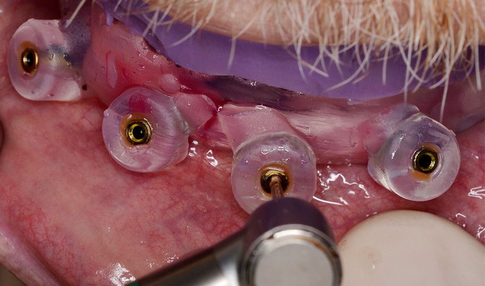
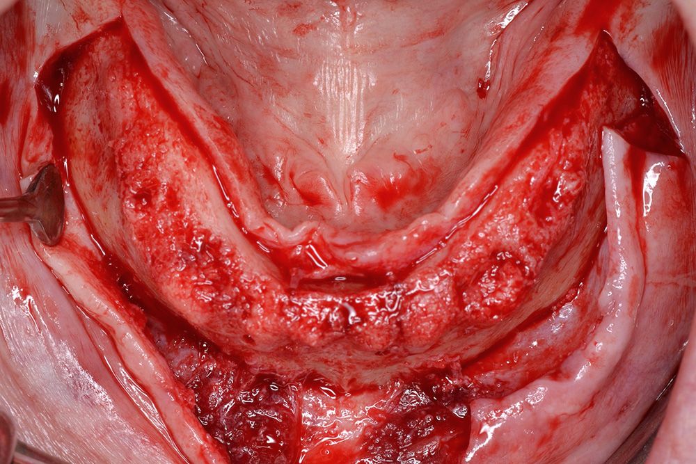
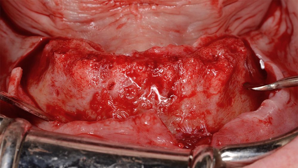
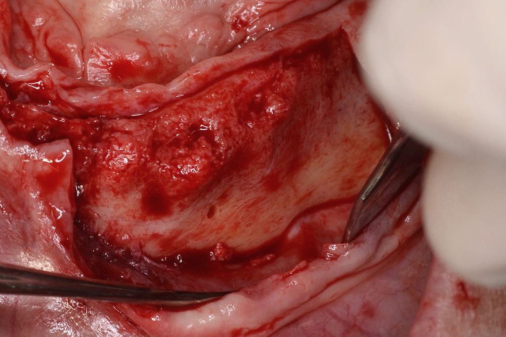
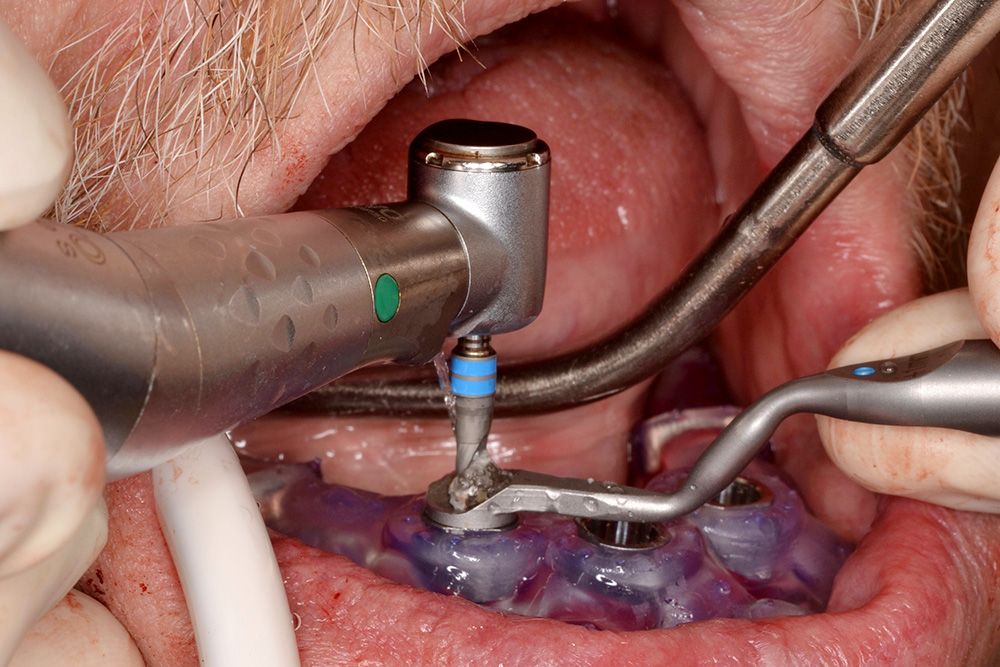
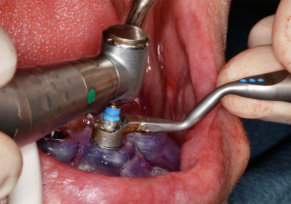
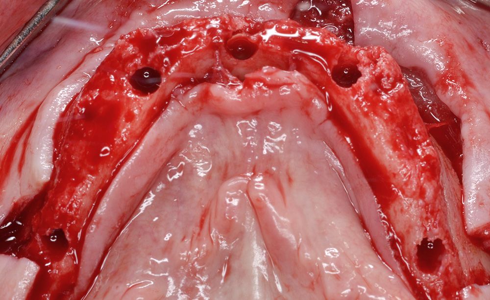
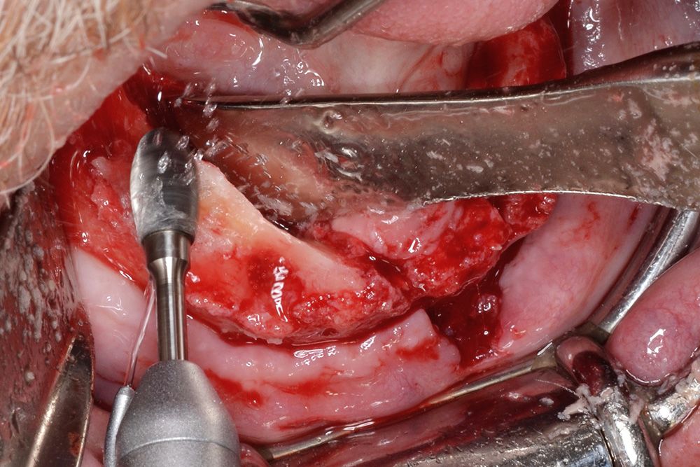
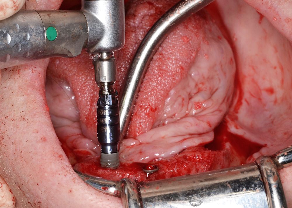
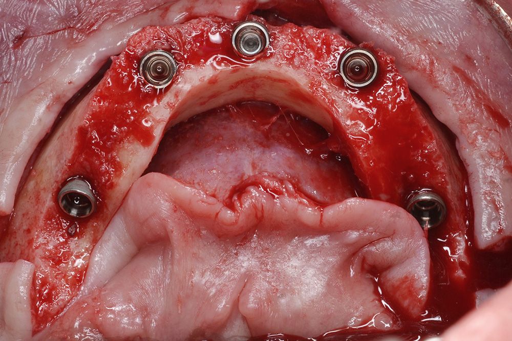
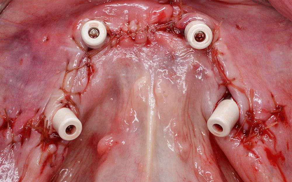
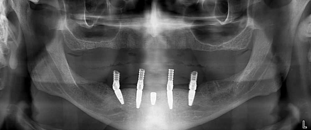
Bilateral local anesthesia was administered. The vertical dimension of occlusion was measured extraorally using a Boley gauge with facial landmarks on the patient’s nose and chin. The occlusal fixation guide was positioned opposing the patient’s maxillary conventional denture and verified with polyvinyl siloxane bite registration (Figs. 9-12). While the patient was in centric occlusion, four 1.3mm diameter osteotomies were created in the labial plate through the alveolar mucosa using the Straumann® template fixation drill (Figs. 13-14). The surgical guide was removed, and crestal incisions were made bilaterally extending to the external oblique ridge. Distal releasing incisions and an anterior releasing incision 4mm to the left of the midline were made (Figs. 15-16). After full-thickness flap reflection, the mental foramina and neuromuscular bundles were visualized and isolated (Fig. 17). The surgical placement guide was inserted and stabilized using four 1.3mm diameter Straumann® template fixation pins (Fig. 18). Sequential osteotomy preparation was completed to depth through the Straumann® 5mm T-sleeves at each site using the Straumann® Bone Level Tapered (BLT) fully guided surgical kit (Figs. 19-21). Following removal of the surgical placement guide, approximately 5mm of alveoloplasty was completed (Fig. 22). Restorative space, osteotomy depth, and avoidance of vital anatomical structures were verified. Two Straumann® BLT SLActive® (RC, 4.1mm diameter, 10mm) implants were placed at sites #35 and #45. Two Straumann® BLT SLActive® (RC, 4.1mm diameter, 12mm) implants were placed at sites #32 and #42, and a single Straumann® BLT SLActive® (RC, 4.1mm diameter, 8mm) implant was placed at site #31 (Figs. 23-24). Straumann® angled screw-retained abutments were torqued to 15Ncm, and sites with thin labial bone over the implant were grafted with autogenous bone as needed. Flaps were repositioned and sutured with 4-0 chromic gut (Figs. 25-26).
Restoration procedure
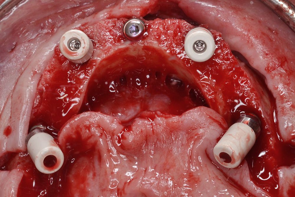
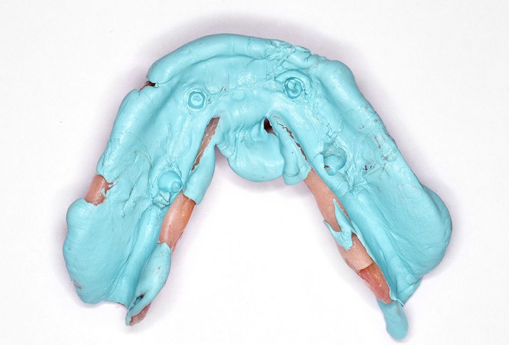
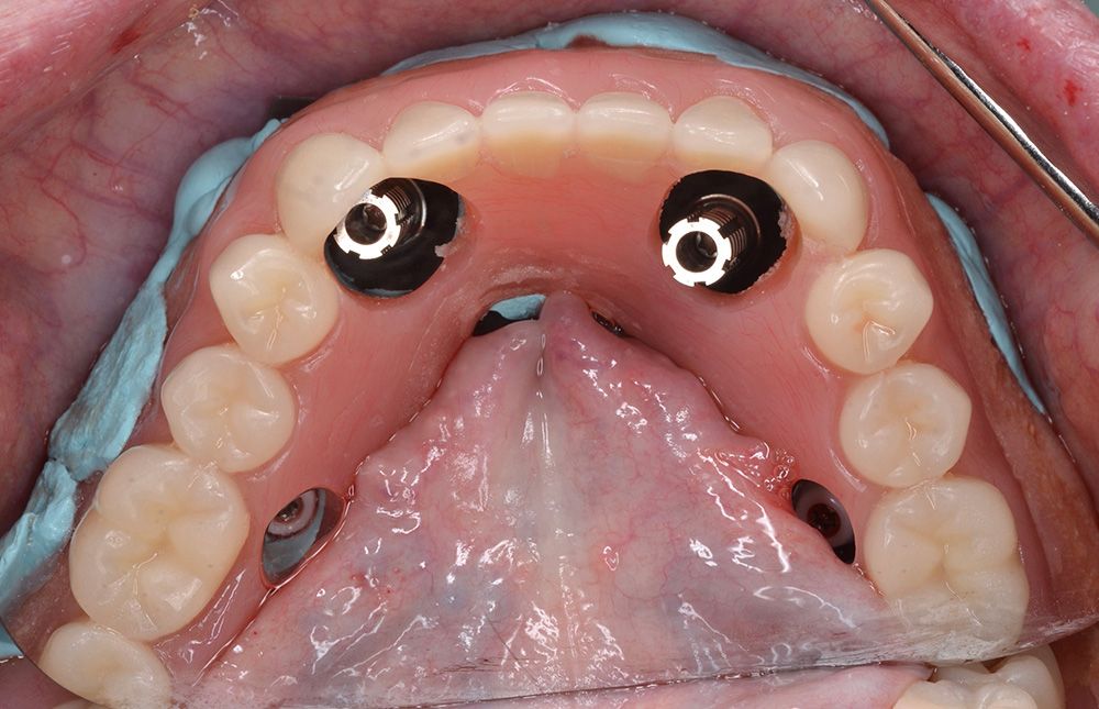
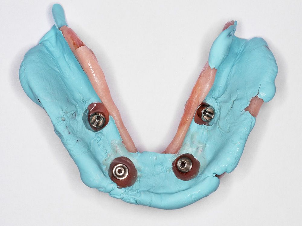
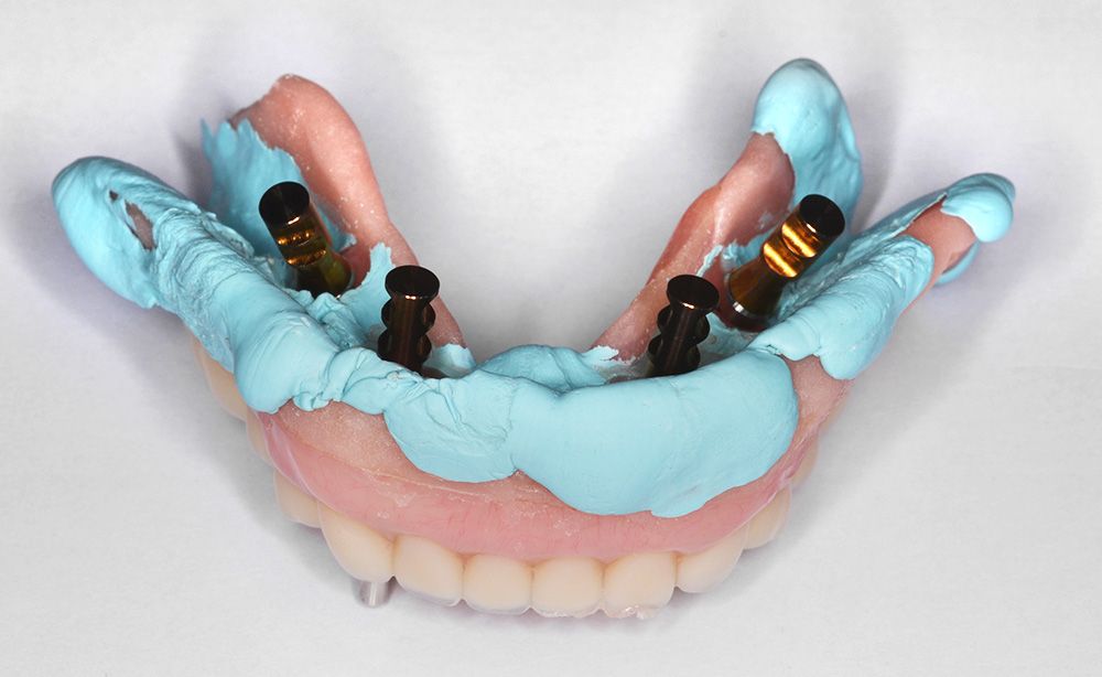
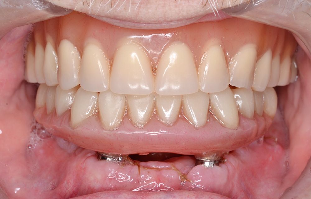
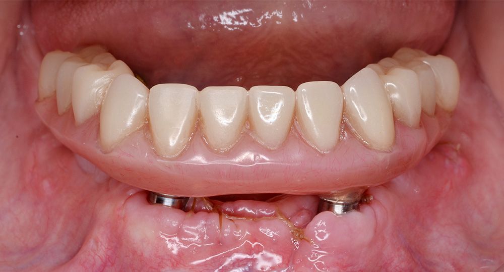
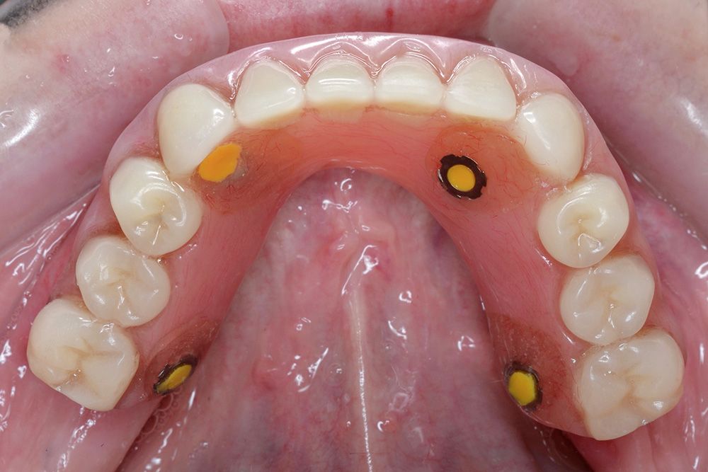
After implant placement, the mandibular complete denture was re-inserted with fast-set PVS material placed on the intaglio surface to indicate implant location (Figs. 27-30). Space was made for the temporary copings, healing caps were removed, and Straumann® RC temporary copings were placed on implants #35, 32, 42, and #45 (Figs. 31-33). Copings were luted to the conventional denture using cold-cure pink acrylic resin. The prosthesis was converted from the pre-existing conventional complete denture to an interim fixed detachable prosthesis by reducing the flanges and providing adequate hygiene space. Occlusal screws were torqued to 15 Ncm, and occlusion was adjusted to have uniform contacts in centric relation and balanced occlusion in excursive movements. The screw access holes were filled with extra light viscosity PVS material (Figs. 34-36). A post-operative radiograph was taken after delivery of the prosthesis (Fig. 37).
Outcome and conclusions
Utilization of the template fixation pins was an essential step in transitioning the mucosal borne occlusal guide to a secure, bone-supported guide. The pins are designed to ensure the digitally planned guide is in the correct surgical position to provide a restoratively driven outcome. In this case, the virtual planning models and the actual outcome demonstrated that the Straumann® Guided Surgery System provides a high level of precision for the purposes of implant positioning.


