Immediate Full arch reconstruction of the edentulous mandible using the Straumann Pro Arch Concept
A clinical case report by Anand Krishnamurthy, India
Introduction
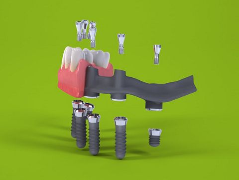
This documented clinical case describes the successful rehabilitation of an edentulous mandible using the Straumann Pro Arch Concept for an immediate Interim hybrid prosthesis, and utilizes the BLT implants in conjunction with the angulated screw retained abutments (SRA) for appropriate prosthetic angulation correction, combined with immediate loading on the implants. The concept of Straumann® Pro Arch is a fixed rehabilitation, which encompasses the whole procedure from removal of hopeless teeth, immediate placement and immediate loading of the implants, tailored to individual patient. It also includes treatment planning before the surgery and converting the temporary bridge to the final full-arch prosthesis afterwards.
The Straumann® Pro Arch solution comprising of the implants, screw-retained abutments and screw-retained prosthesis has become the choice to meet the patient's functional and aesthetic goals. It provides the flexibility required in a situation with limited anatomical structure of a deficient jaw bone volume, where conventional treatment may not be feasible. The inherent flexibility in the Straumann Pro Arch process proved to be beneficial for the following reasons :
1) The surgical planning to identify the appropriate position of the dental implants to optimize primary stability during placement and to minimize the need for any augmentation procedures and also to avoid the proximity to important anatomical landmarks like the inferior alveolar nerve and the mental foramen.
2) The transitional phase allowing for the conversion to a dental implant supported fixed hybrid prosthesis working with the Straumann Screw Retained Abutments (SRA) to account for the angled position of the dental implants. This makes the entire concept prosthetically driven right from the start.
Initial situation
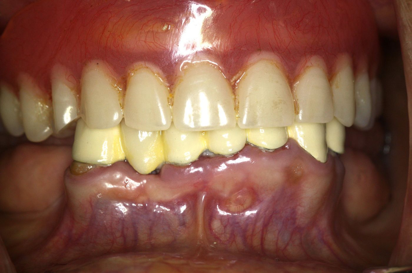
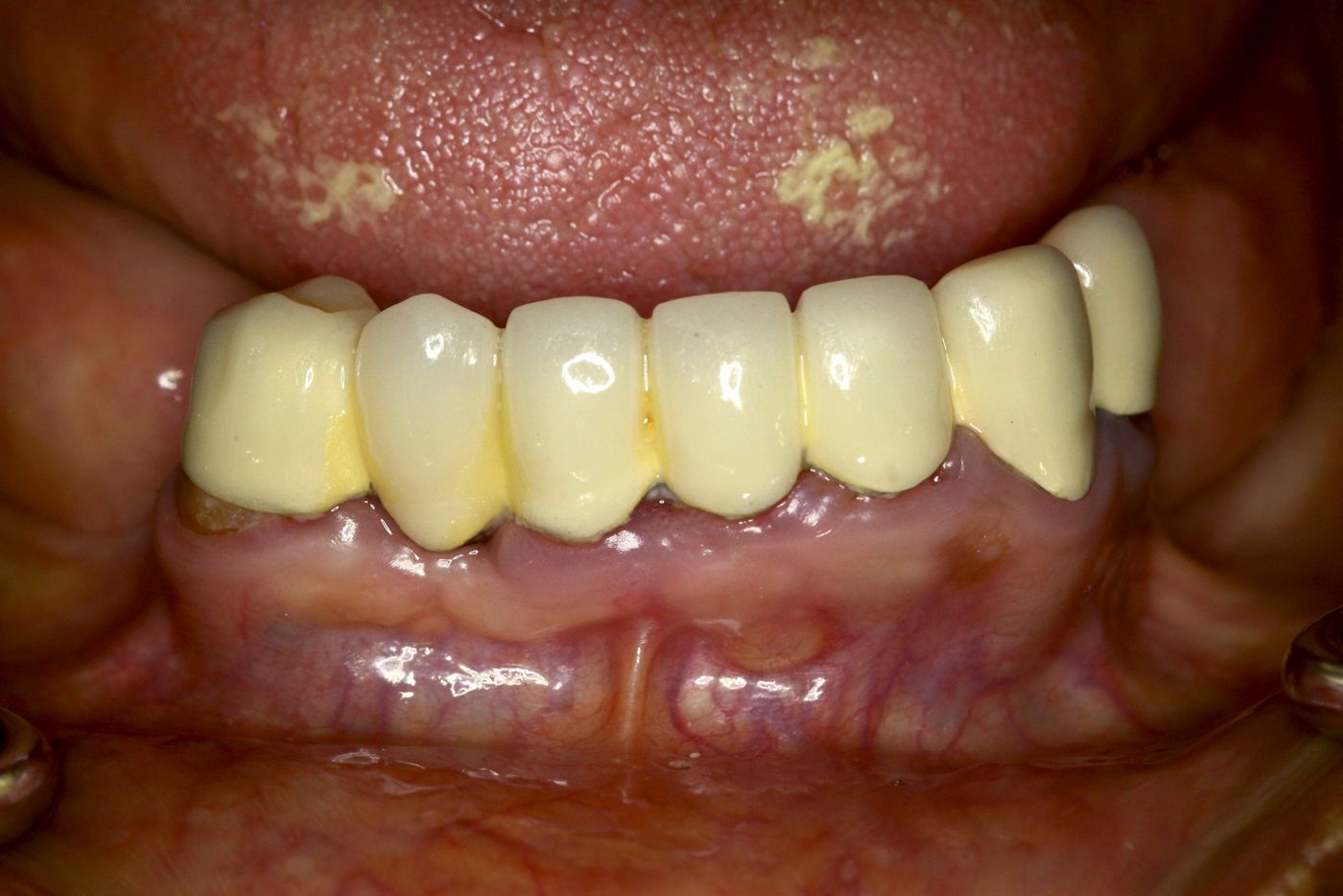
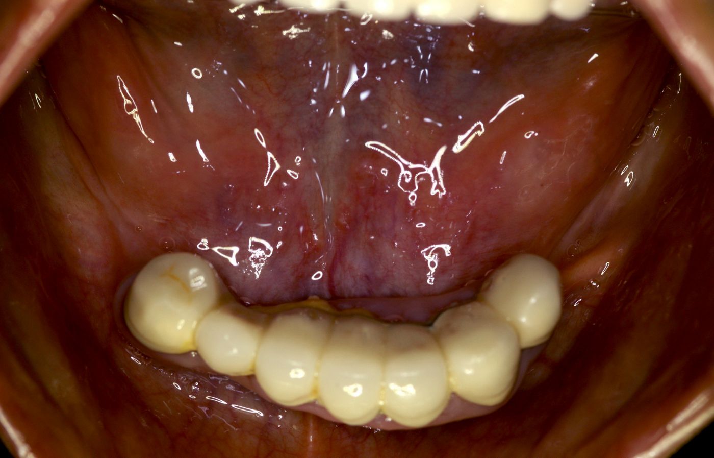
The patient was a 65 year-old female, non smoker, non diabetic with no relevant medical history or any major ailments. She was an existing denture wearer in the upper jaw and presented to us with a failing 7 unit crown and bridge work in the lower jaw supported on teeth # 33,34, and 43 (Figs. 1-3)
The existing bridge was clinically mobile and the abutment teeth were tender and clinically symptomatic. The patient expressed her discomfort and inability to chew food due to her existing lower bridge and desired a fixed restoration which would be functional and also aesthetic.
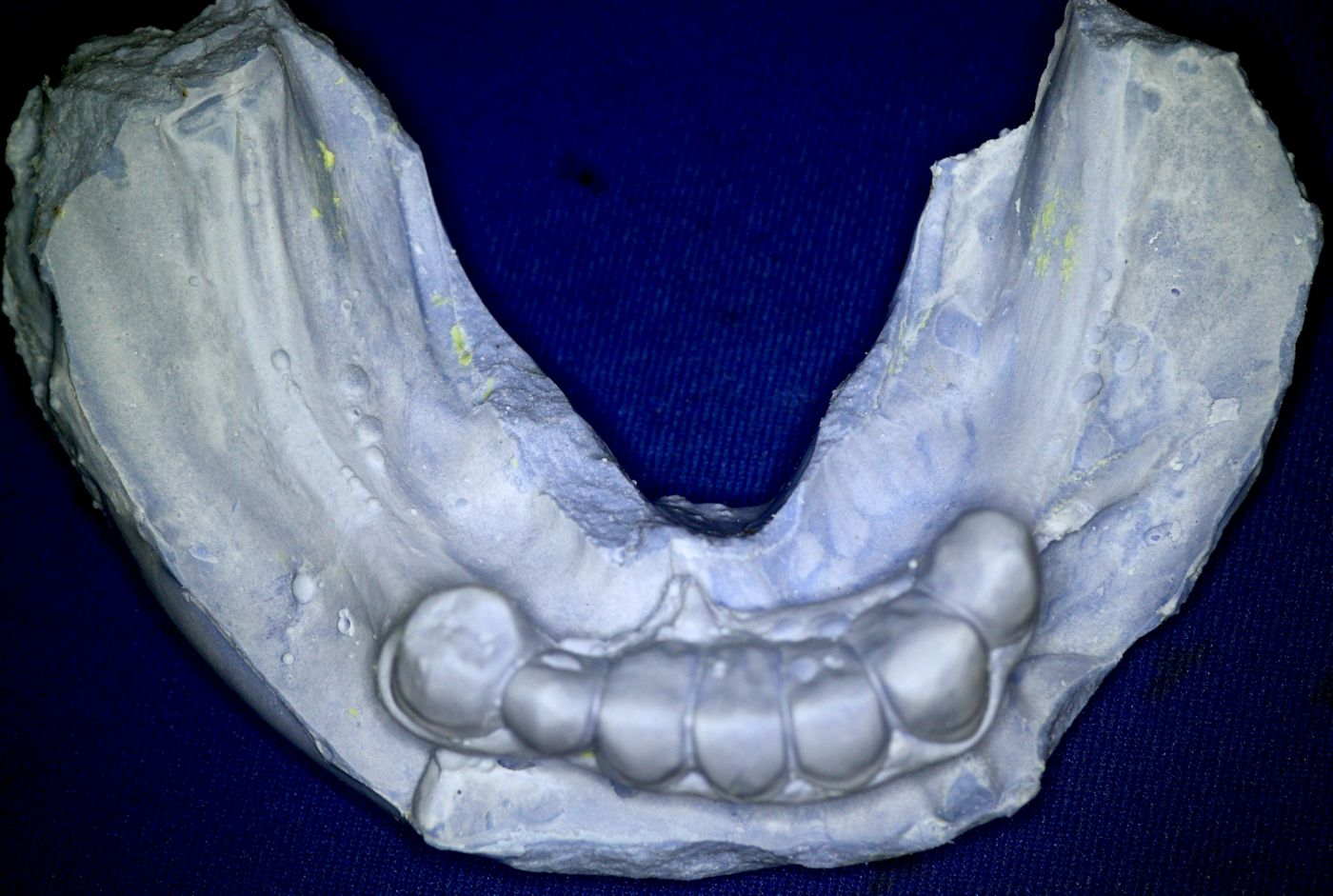
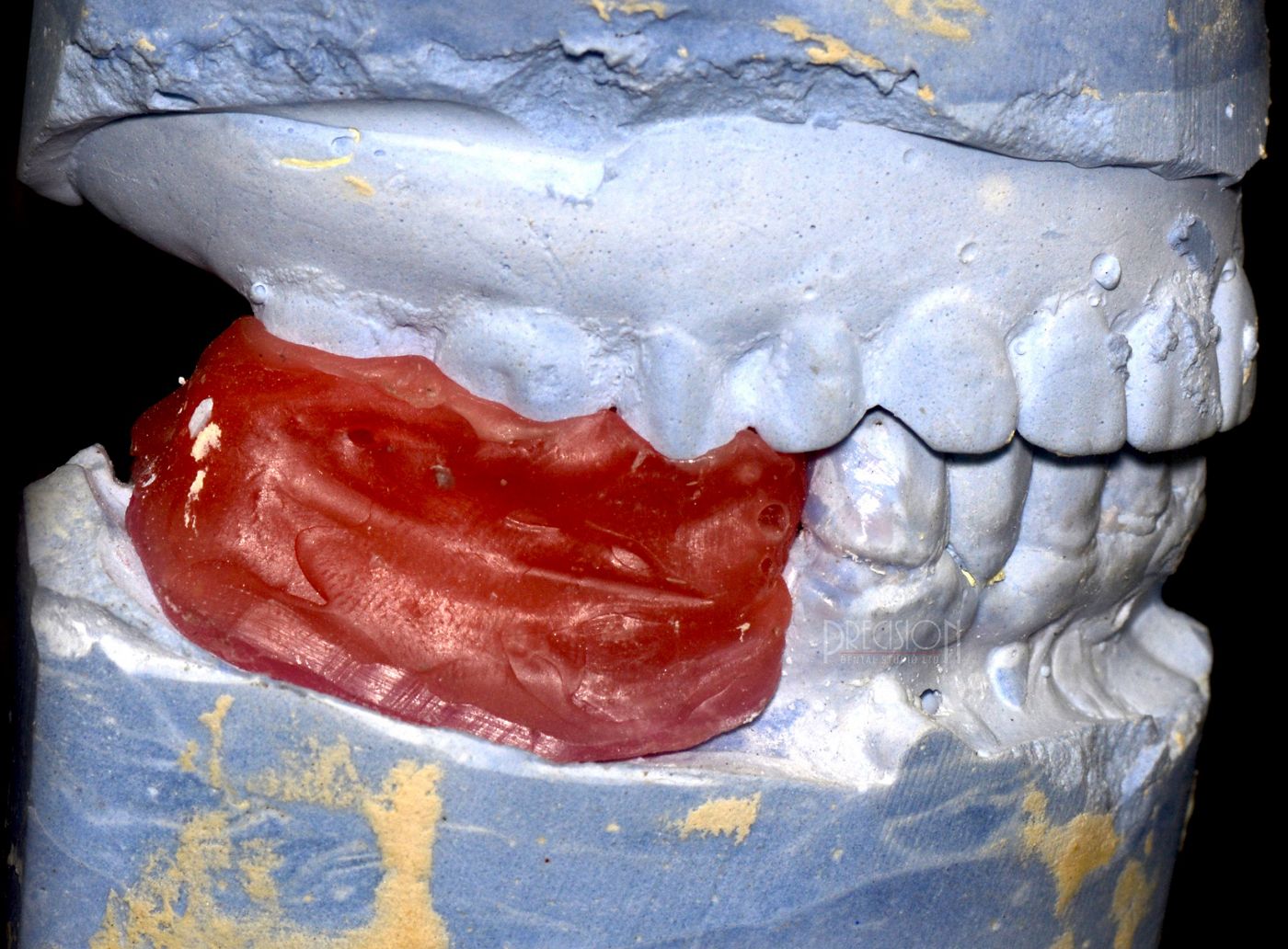
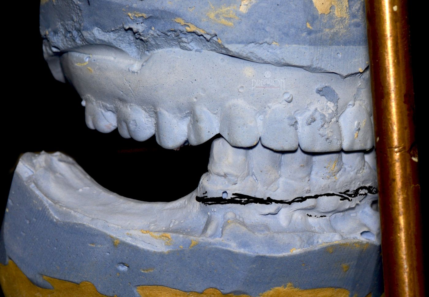
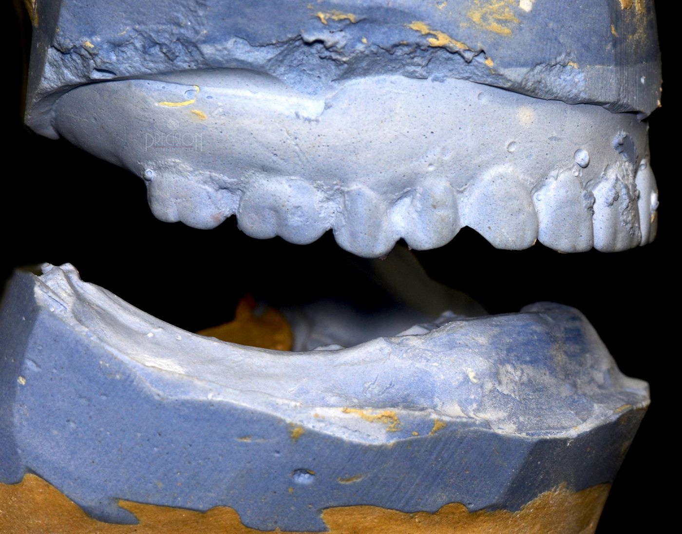
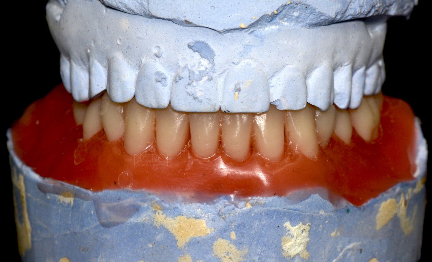
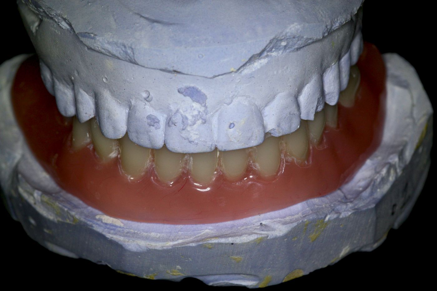
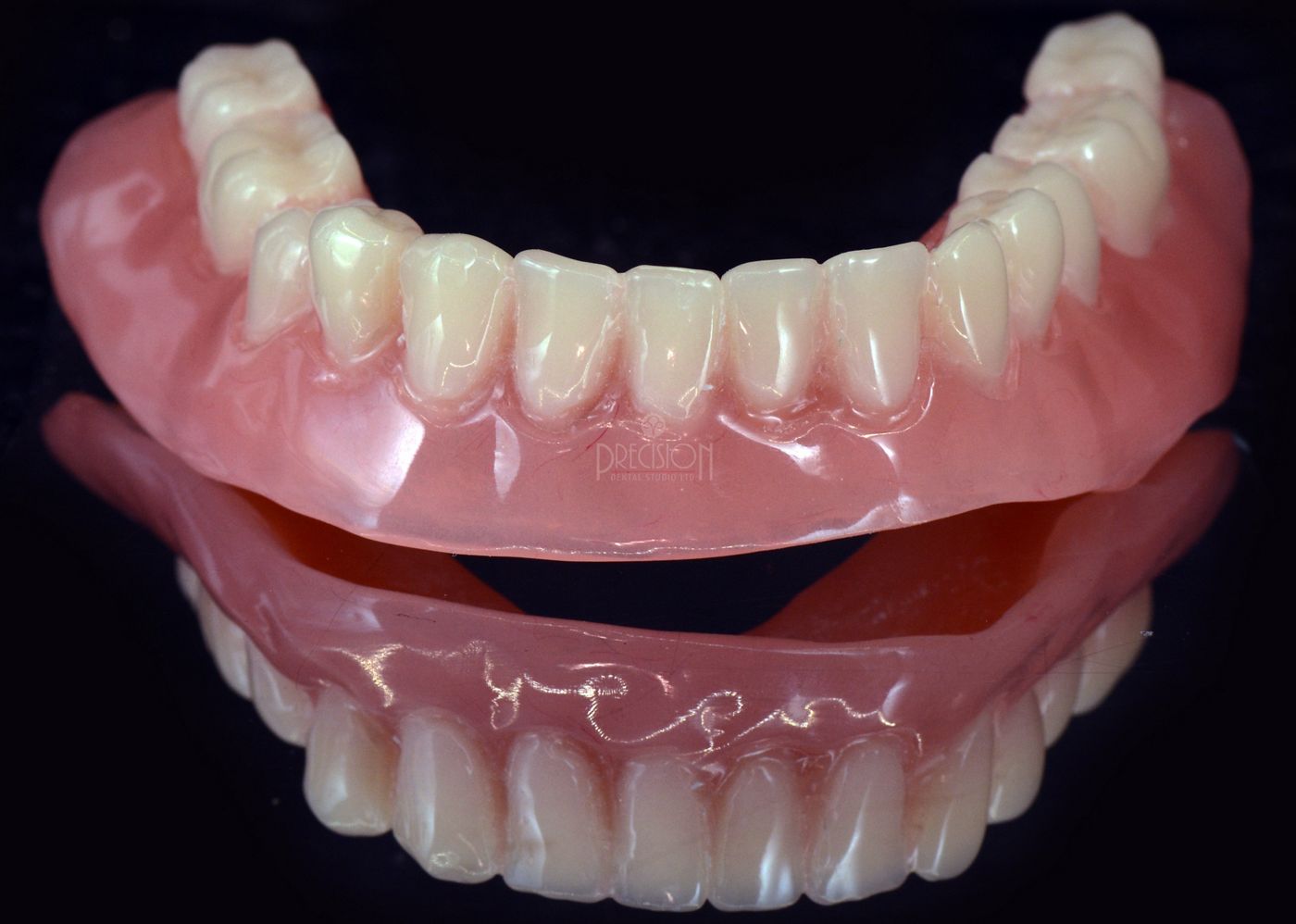
Study models were created and mounted in articulation at the existing vertical dimension, which was appropriate enough to proceed with the proposed treatment plan as there was no loss of vertical height or a collapse (Figs. 4-6). The study models were also duplicated to fabricate a diagnostic wax up for the lower jaw at the centric relation to outline the teeth set up for the immediate provisional prosthesis (Figs. 7-8).
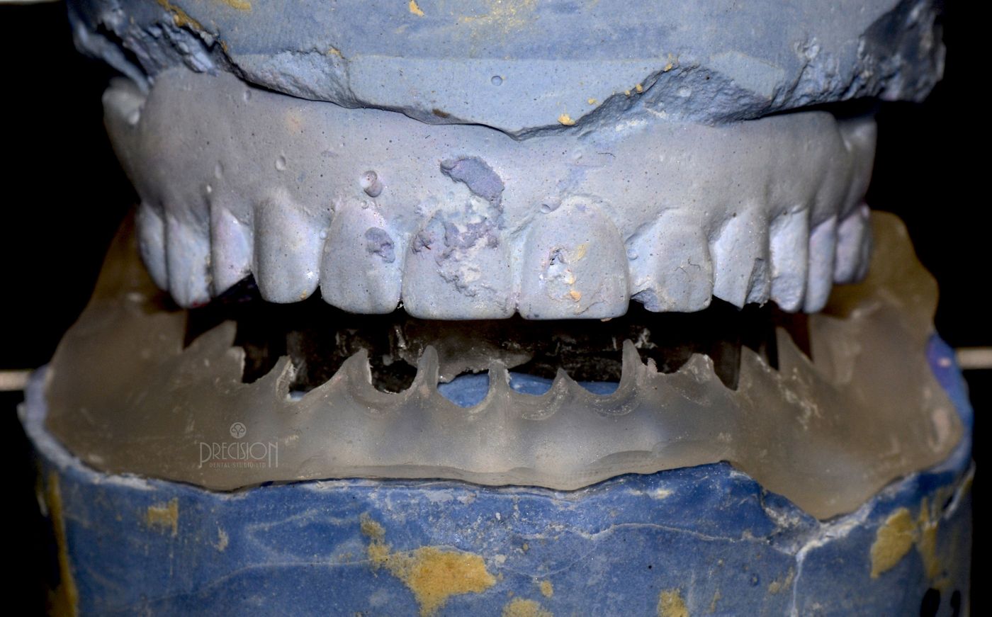
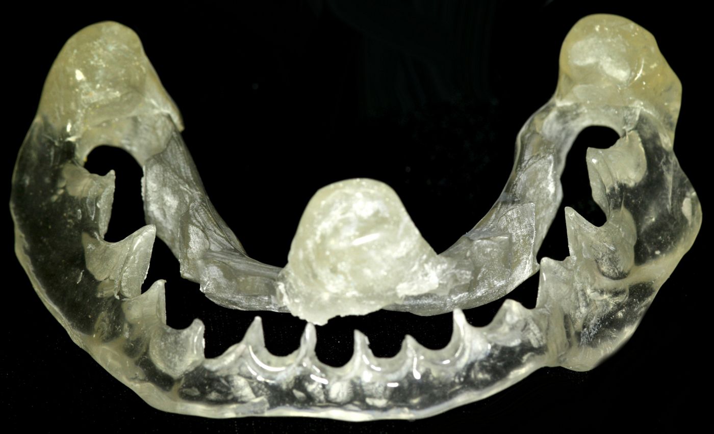
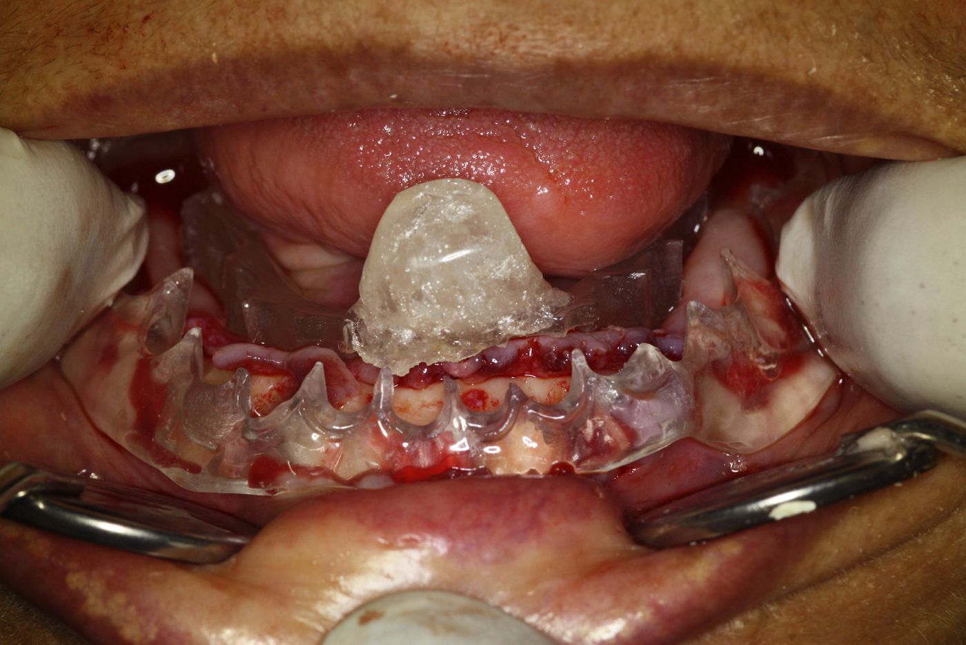
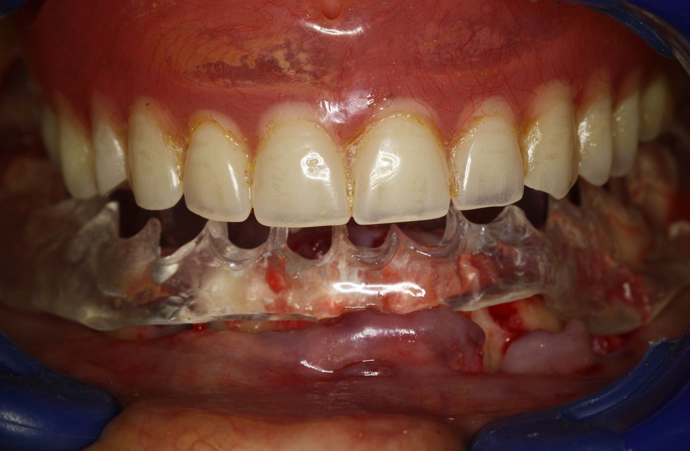
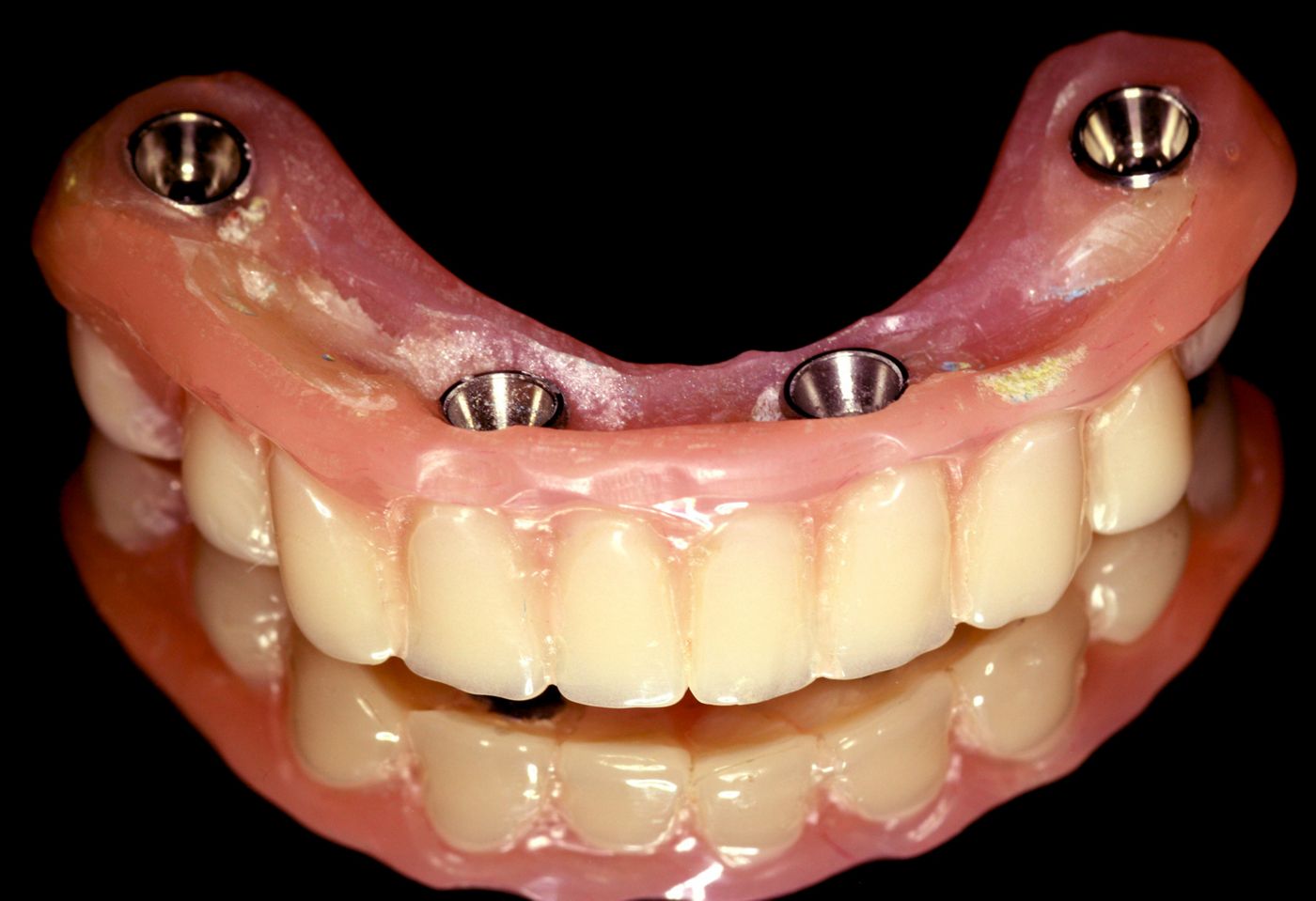
The immediate provisional prosthesis fabricated by the lab was similar to an acrylic complete denture with acrylic teeth, which would be converted chair-side into a screw retained fixed prosthesis during the surgery and connected to the implants after the surgery. The lab was also instructed to create a clear resin prosthetic index (Figs. 9-12) as per the proposed teeth set up at the same vertical relation. Acrylic vertical stops were also added in the index to verify the amount of crestal bone to be removed during the surgery to create ambient space for the SRA, the coping height and the teeth set up above them.
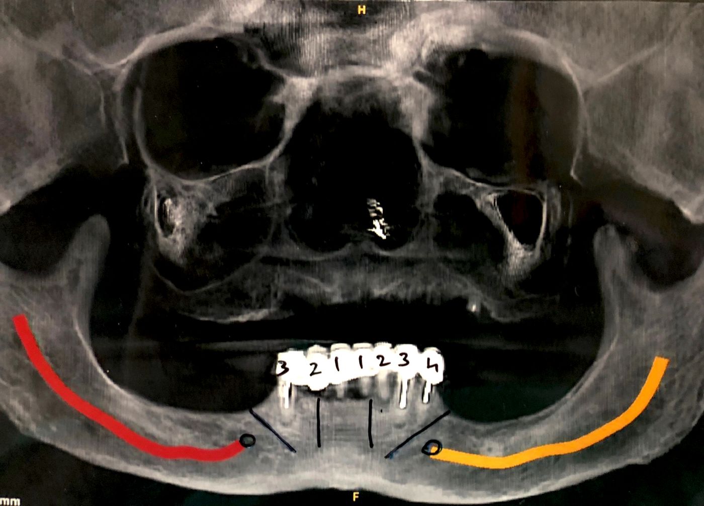
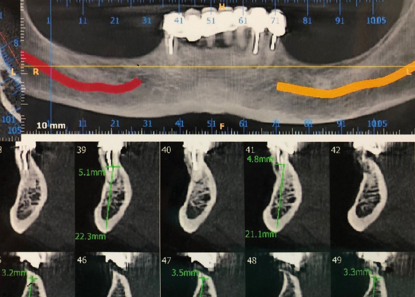
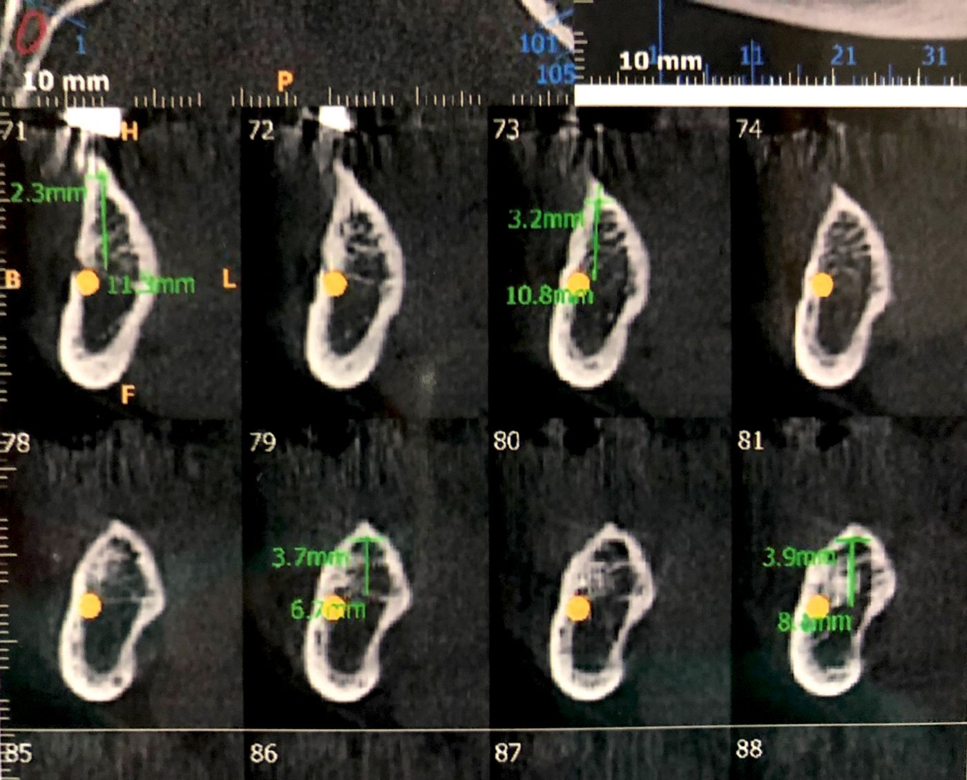
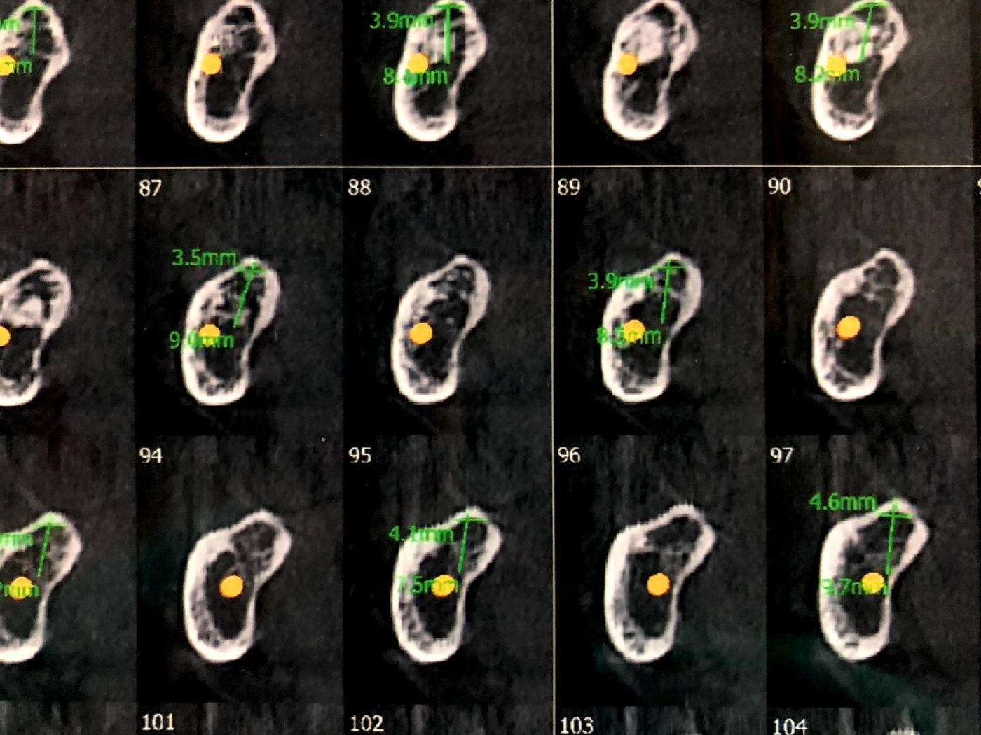
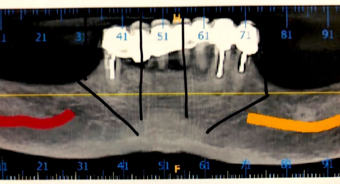
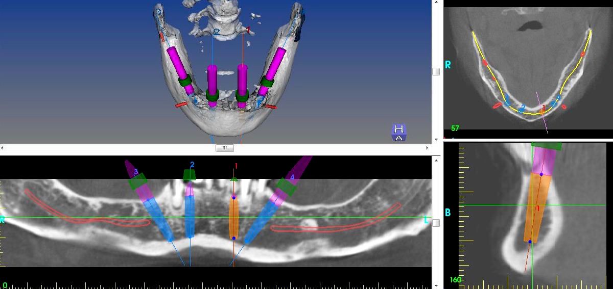
Radiographic evaluation was done using a CBCT Scan of the lower jaw to consider the bone volume available for placements of four BLT implants and the DICOM data was transferred to a implant planning software to create a virtual 3D surgical plan for the patient. The Scan and the 3D analysis indicated only sufficient bone height and width available in the Inter-foramina area and beyond the foramen distally there was deficient bone height as seen the images. The correlation of the 3D planning with the prosthetic index was important to determine the position of the distal most implants to avoid any cantilever of teeth beyond the abutment trajectory (Figs. A, B, C, D, E, F).
Treatment planning
The treatment plan was to realize the Pro Arch concept in the lower jaw with immediate temporization using a provisional hybrid screw retained prosthesis. Two Straumann BLT Roxolid SLActive implants with ∅ 4.1 mm, 14 mm length were placed in anterior region and two 16 mm length in the posterior region, based on our 3D software simulation planning. Two distal 16 mm implants were tilted to gain A-P spread and bypass the mental foremen to reach out more posteriorly in abutment trajectory as per the proposed teeth set up. The entire treatment plan was restoration driven, as the prosthetic teeth positions determined the implant positions and the amount of tilted angulation needed on the distal implants was calculated to achieve the same prosthetic goal without any cantilever. The tilting the distal implants was planned to avoid utilizing the bone distal to the mental foramen, which would have needed bone augmentation otherwise due to the deficiency.
The planned implant placement modality was type 1 (immediate, post-extraction) and the planned loading modality was immediate loading. An open flap approach was planned as there was a need to perform crestotomy after the extraction of hopeless teeth. The Implant osteotomies were to be performed free hand with the aid of the prosthetic guide to help us in locating the exact positions of the implants in relation to the prosthetic teeth set up.
Lab staff received the treatment planning in preparation for surgery beforehand.
Surgical procedure
Bilateral local anaesthesia was used in the mandible for complete haemostasis during the procedure. The patient was put on an antiseptic Chlorhexidine mouthwash one week prior to the surgery and a prophylactic antibiotic Amoxycillin Clavulanic acid 625 mg BD one day prior to the surgery, which was to be continued for 5 days after the surgery.
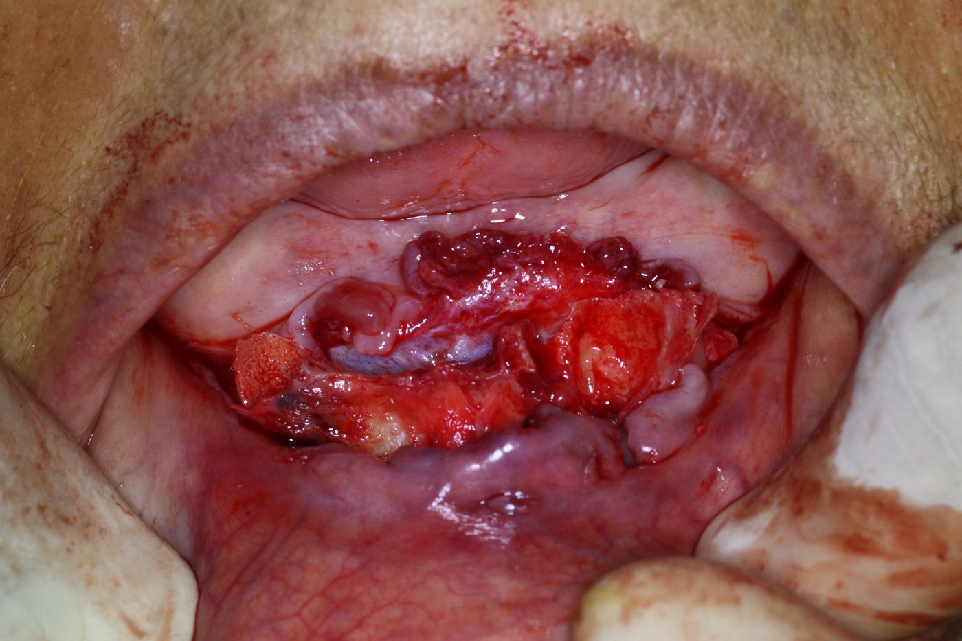
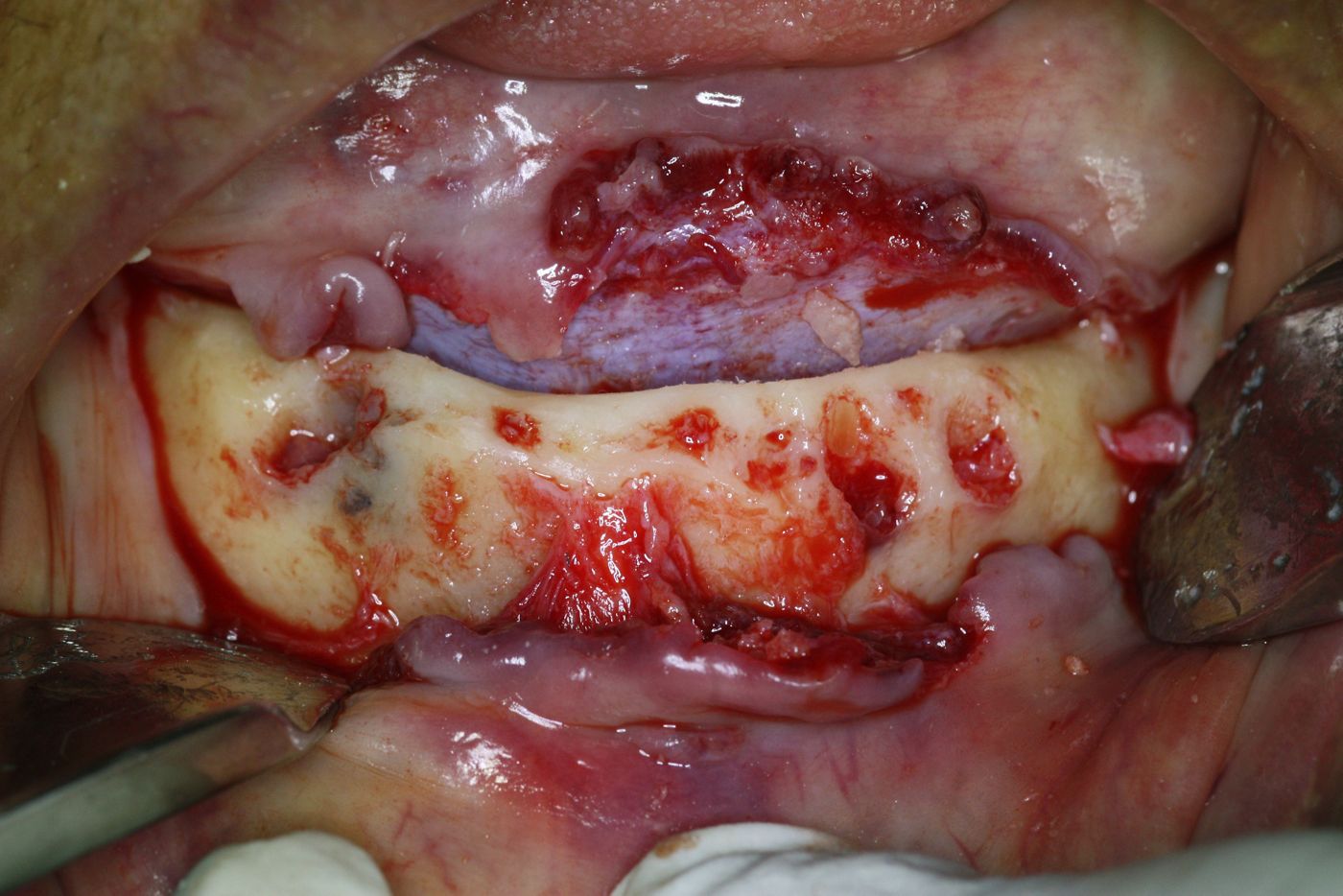
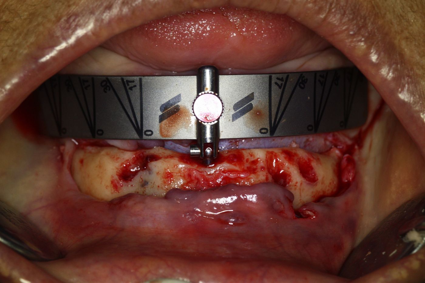
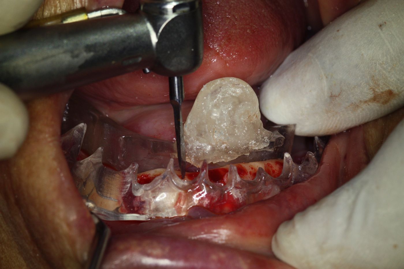
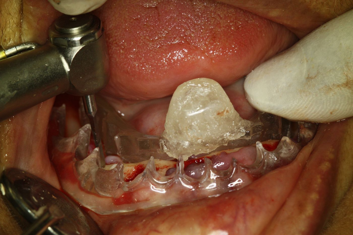
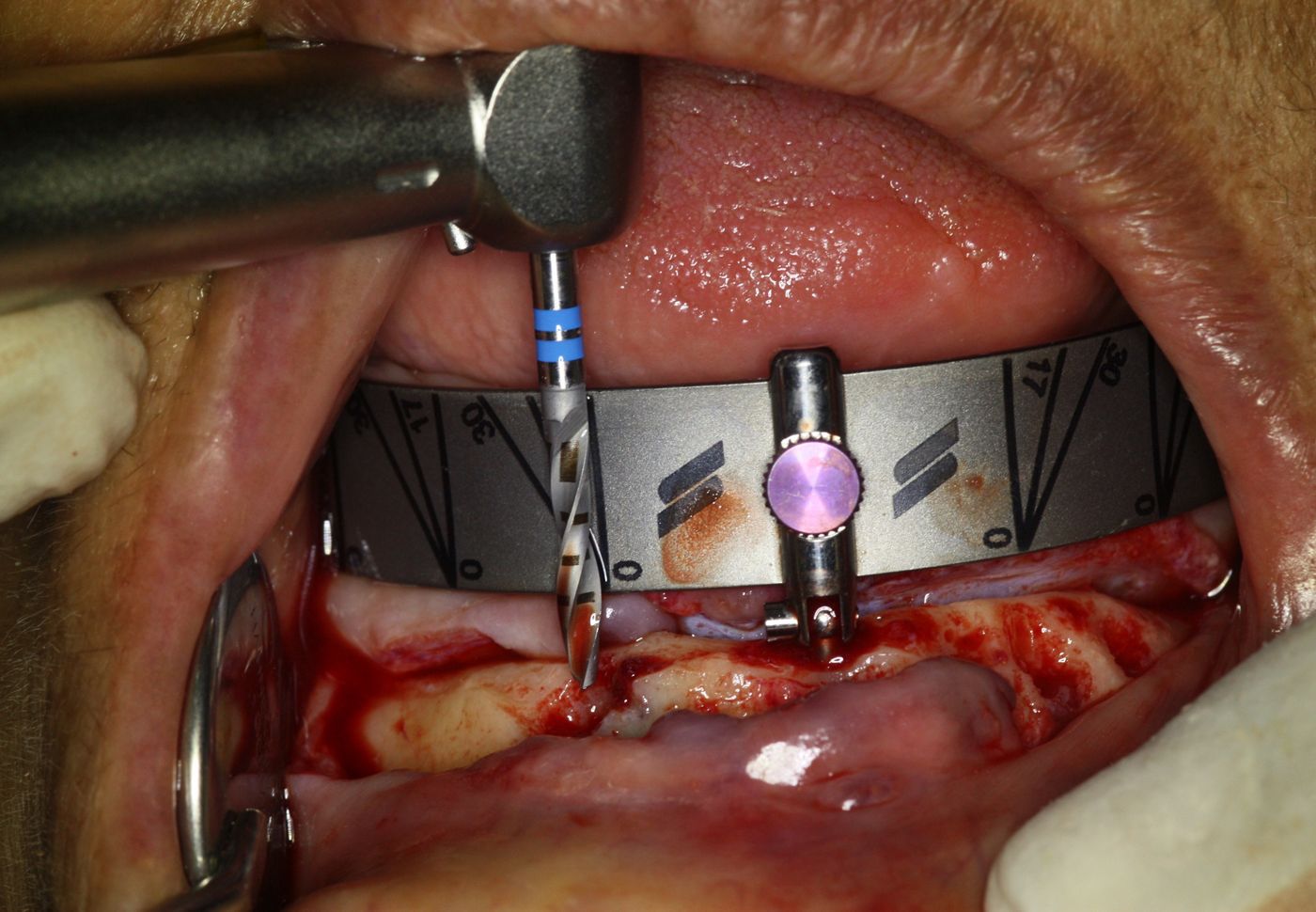
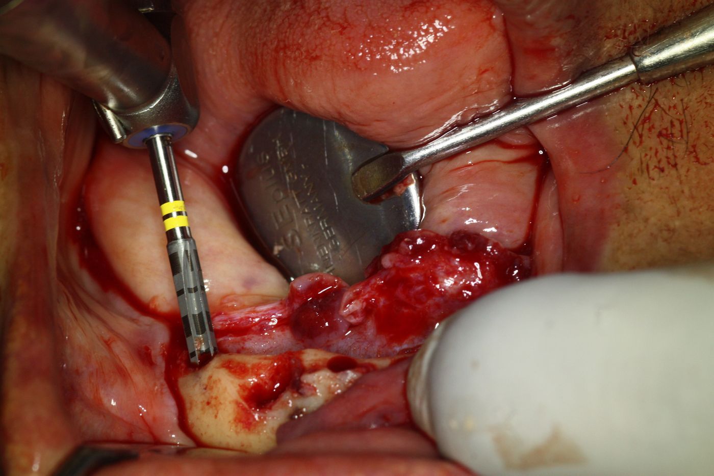
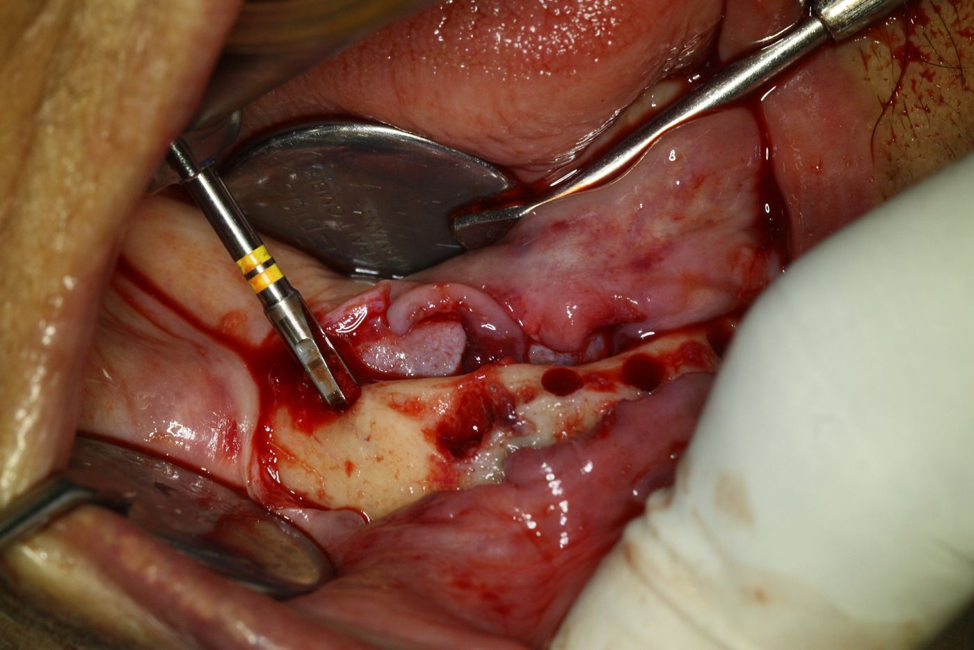
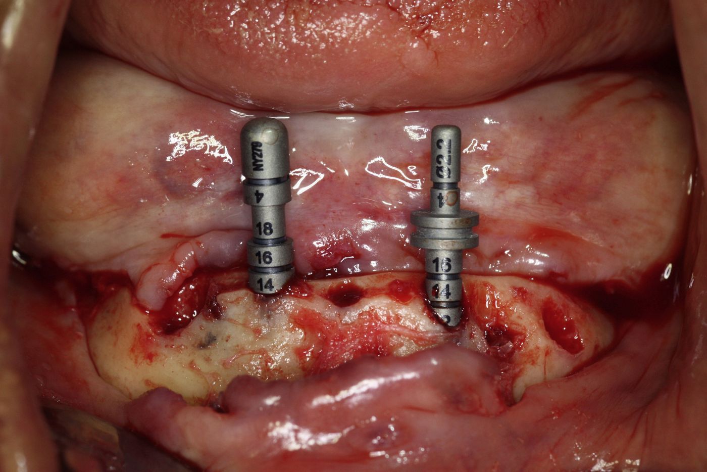
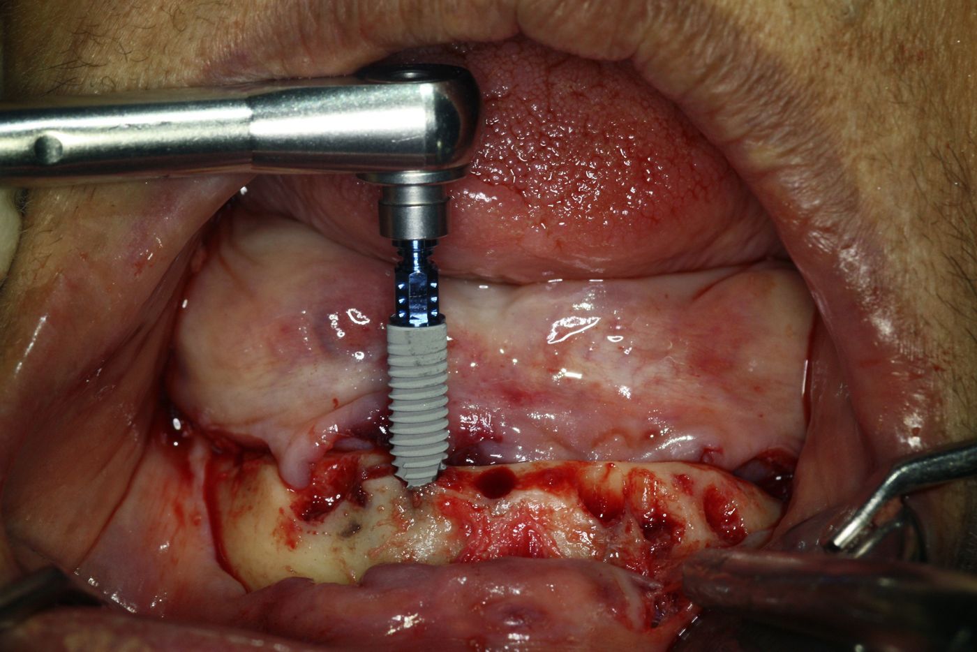
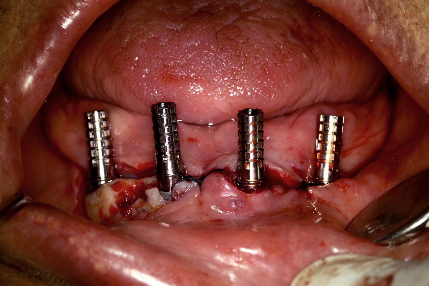
The mobile lower bridge was easily removed attached to the existing root fragments and complete curettage of the sockets was performed. A full thickness flap was raised after that to expose the bony contours and all the sharp areas around the sockets were eliminated (Fig. 13). The Prosthetic index was kept in place in relationship to the upper denture in centric relation to evaluate the amount of bone vertical reduction or crestotomy needed. The idea was to gain a total vertical space of 10-12 mm above the desired bone level to accommodate the space needed for SRA, the coping and the teeth set up. Accordingly the bony crest was flattened with a rose-head drill with irrigation until we achieved the goal (Figs. 14-16). The flattening of the ridge also helped establishing a good flat platform to start our implant osteotomies using the prosthetic guide as a surgical marker, which indicates prosthetically ideal position of the implants. The purchase points of four implant osteotomies were marked through the prosthetic index in the position of # 32, 35, 42 and 45 (Figs. 18-19). An additional purchase point was created in the mid centre of the mandible between the 31 and 41 regions to facilitate the positioning of the the Straumann Pro Arch guide (Figs. 17-20). The template has fixed laser markings with set angulations of 0 degree, 17 degree and 30 degree. It can be bent according the individual arch form and moved mesiodistally to match the purchase points created. The anterior two implant osteotomies were performed using the 0 degree angle and the distal two implant osteotomies were performed using the 30 degree angle, thereby bypassing the mental foramen safely (Figs. 21-22). The standard Straumann drilling protocol was followed to place two BLT Roxolid SLActive ∅ 4.1 mm, 14 mm length implants anteriorly and 2 BLT Roxolid SLActive ∅ 4.1 mm, 16 mm length implants posteriorly (Figs. 23-24). Excellent primary stability was achieved in all four implants placed as per the drilling protocol.
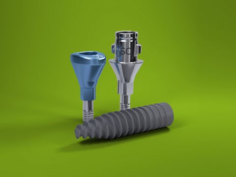
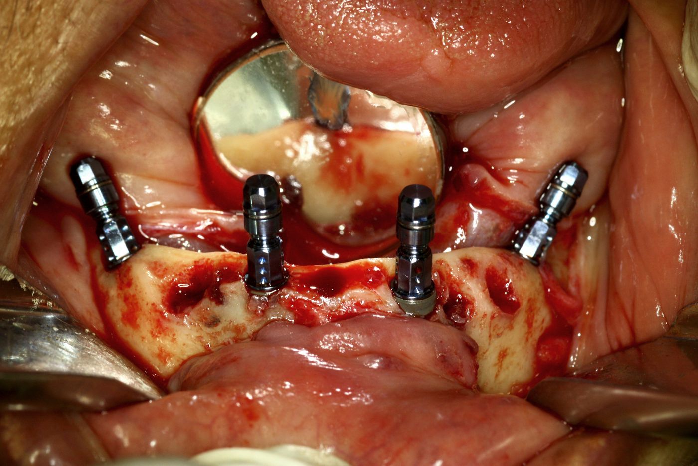
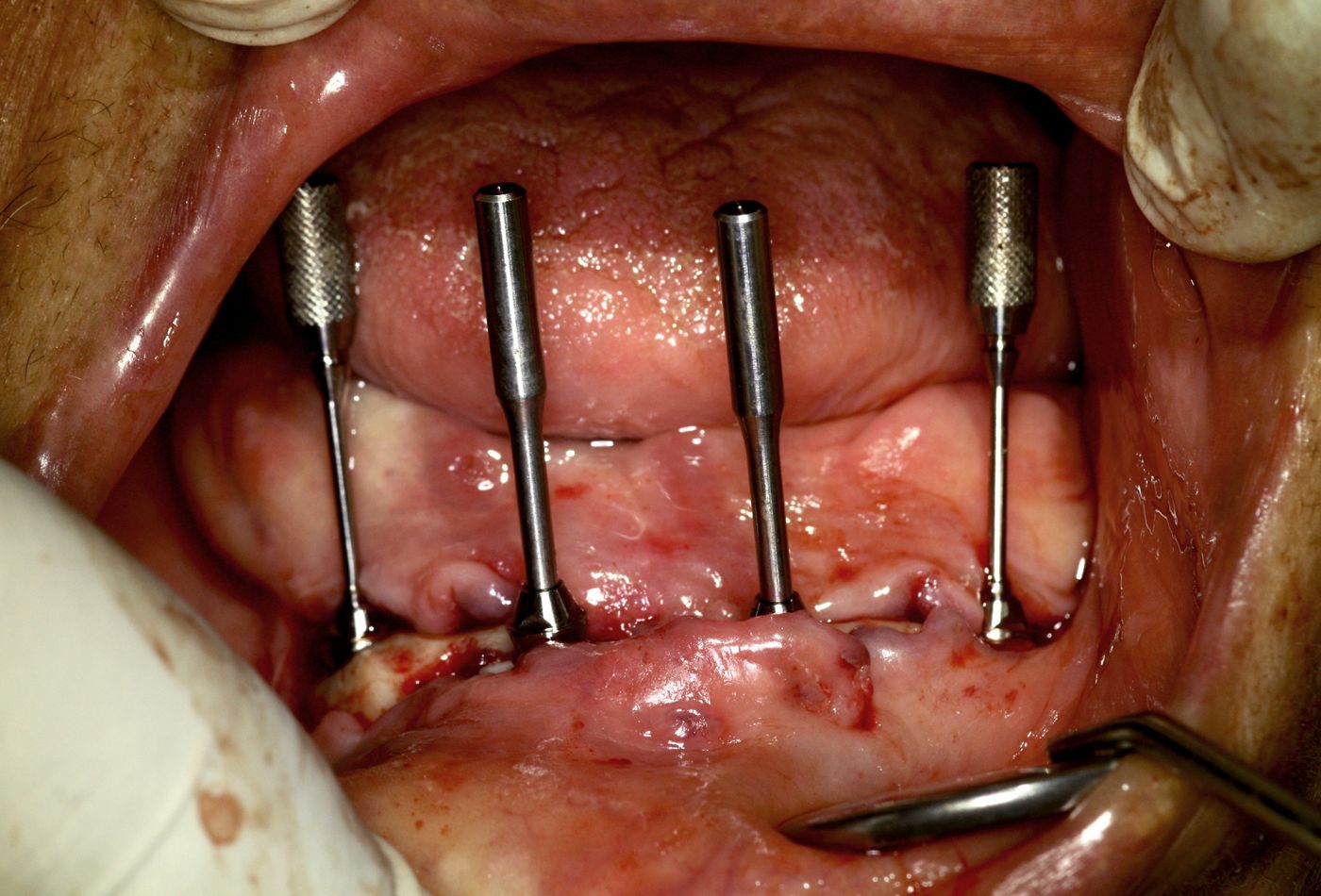
Once all four implants were placed the Loxim attachment served as a guiding tool to verify the position and angulation of the implants (Fig. 25). The excess overhang of bone created due to the tilting of the implants was removed using the RC guiding sleeve and the RC bone profiler 3 drills. This was to ensure that there would be no bony interference in the final seating of the angulated SRA abutments using the transfer and alignment pins. In this case we used two RC straight SRA GH 2.5 mm on the anterior straight implants and two RC 30 degree angled SRA GH 4mm on the posterior tilted implants to bring out the transmucosal emergence. Once all the four transfer and alignment pins are in place they should be exactly parallel to each other (Fig. 26). If not, the tilted distal implants should have been rotated and re-aligned in accordance with the Crossfit connection positions to bring it to more favorable index position for the parallelism*. This may involve moving the implant attached to the Loxim, clockwise (preferably) or anti clockwise by a few degrees to achieve the absolute parallelism needed, which can be finally verified by the transfer and alignment pin. This exact parallelism is to ensure the proper seating of the non-engaging temporary titanium cylinders and the easy pick up of them into the provisional prosthesis. Once the final index positions of all four SRA abutments were verified, these abutments were fastened with the basal screw to 30 Ncm.
Primary closure of the flap was done with single interrupted sutures, making sure that the SRA profile was transmucosal in emergence, with the cone shaped head projecting supragingivally. Some resection of the soft tissue was needed to create the ideal emergence profile and to eliminate any soft tissue overhangs over the SRA. Afterwards, the RC non-engaging titanium copings (NC/RC Coping for SRA) were connected on top of all SRA and the parallelism was verified again (Fig. 27).
* This re-aligning of the implants can be avoided with two different abutment version (A or B). In order to find the correct abutment version, check the height markings (three dots) on the Loxim Transfer Piece. If the height markings are oriented buccally use A-type abutments. If the height markings are not oriented buccally use B-type abutments.
Prosthetic procedure
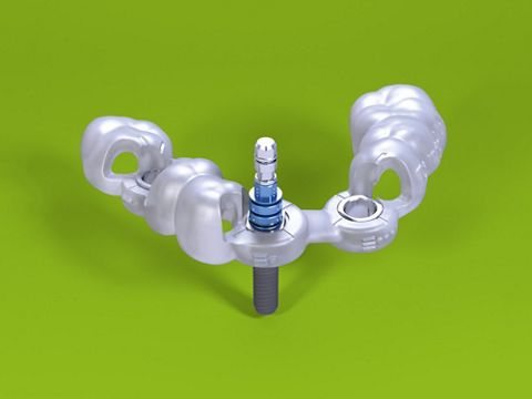
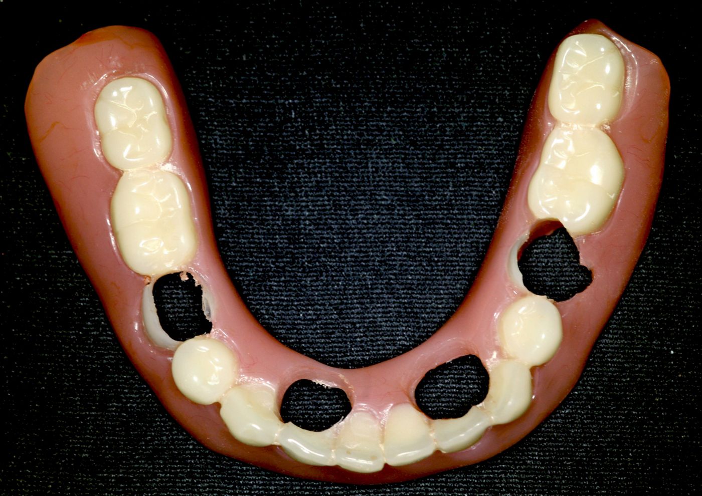
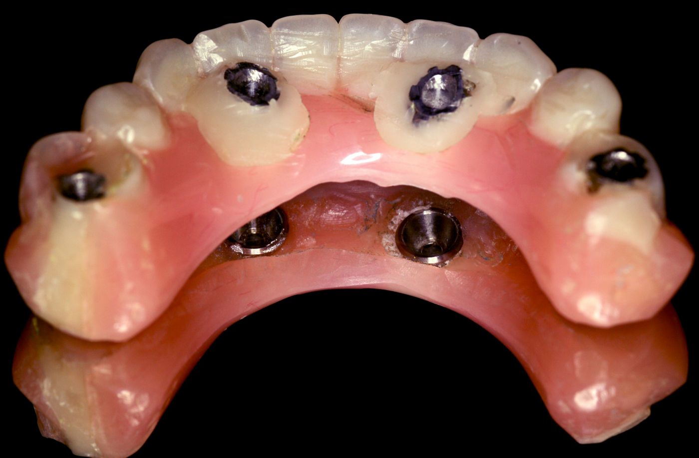
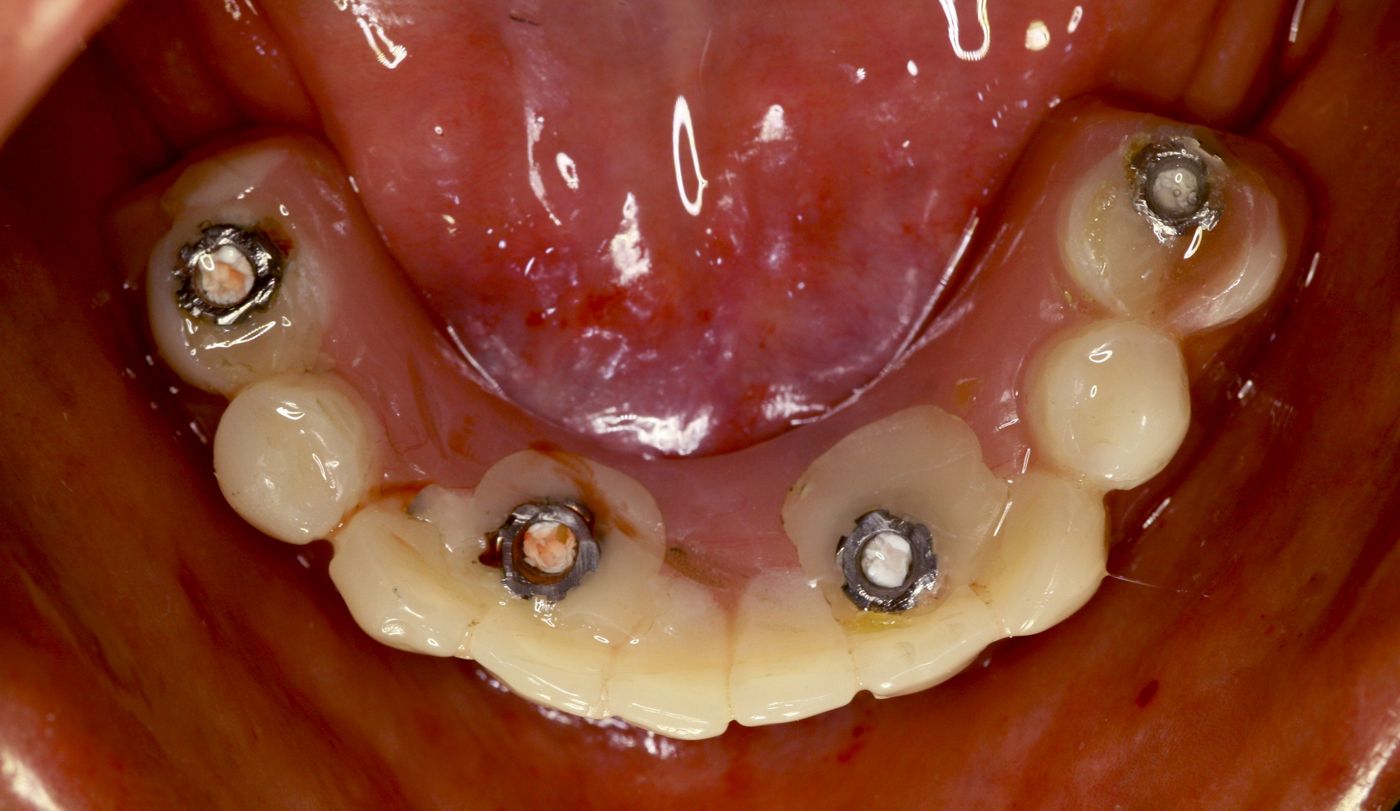
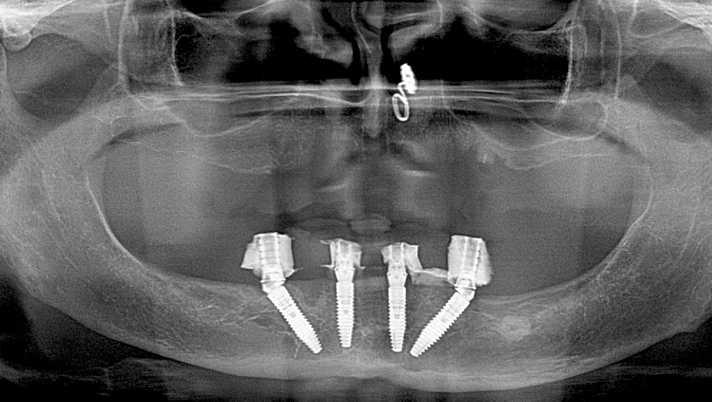
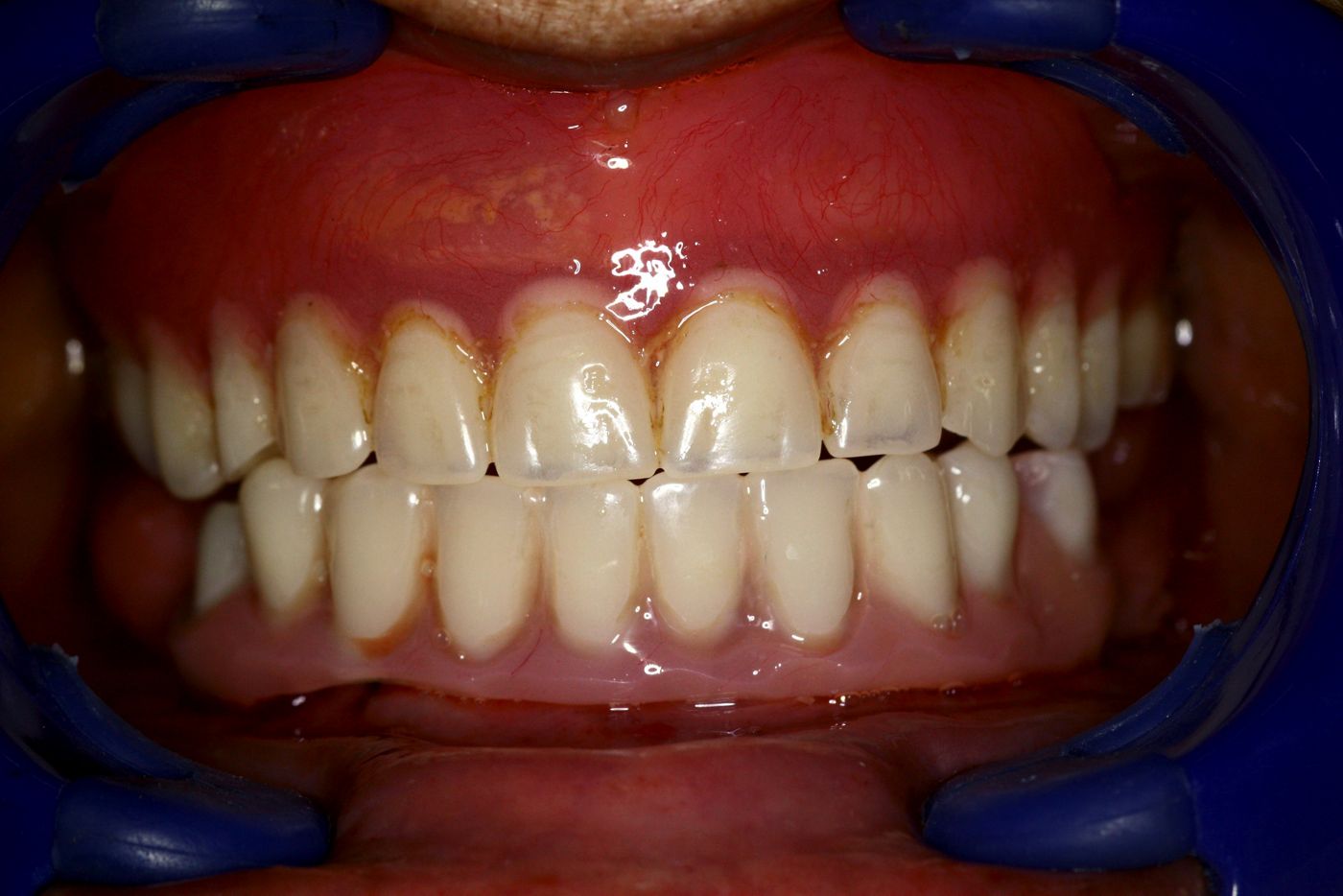
Immediate loading protocol was followed in this case as planned, with immediate long-term provisionalization. The access holes were modified in the acrylic of the prefabricated immediate prosthesis around the positions of # 32i, 35i, 42i and 45i. These teeth positions were duplicated exactly in the prosthetic index, which was used for the osteotomies (Fig. 28). Then the immediate denture was tried on the patient’s jaw to check for the emergence and free trajectory of the titanium cylinders through the holes created. Any acrylic interferences were removed, and the excess height of the cylinders trimmed chair-side with a carbide bur. The exact intercuspation of the immediate denture with the opposing arch denture was verified in centric relationship without any acrylic interferences. Once this was done, the top of the titanium copings were blocked with a soft resin to prevent any resin material from entering inside. A self-curing resin was injected onto the interlocking surface of the titanium copings and the denture was held in centric relation with maximum intercuspation until the resin had set hard. Then the occlusal screw of the titanium coping was loosened on all four SRA, so that the denture could be removed from the mouth, with intra-oral pick up of the titanium copings now incorporated in the denture. This converted prosthesis was now trimmed to remove all the flanges, and the undersurface of the prosthesis was adjusted by addition of self-curing resin in the undercut areas (Figs. 29-30). After sufficient time was spent finishing and polishing, the prosthesis was ready to be connected to the SRA at the same visit. The immediate prosthesis was then screwed onto the SRA with a torque to 15 Ncm. The occlusion was verified for centric relation and anterior guidance and minor adjustments were added. The access holes of the prosthesis was then blocked out with a self-curing tooth coloured resin and some light-cured composite resin (Figs. 31-32). An immediate post-operative OPG X-ray was taken to verify the fit and proper seating of the copings onto the SRA. The proximity of the implants in relationship to the anatomical landmarks were also verified with the OPG (fig 33). The patient was instructed on the maintenance and soft diet regime. First recall was within 24-hour time to evaluate for post-operative pain, edema or any other discomfort (Fig. 34).
Treatment outcomes
No complications or adverse situation was observed at any time during the treatment, from the planning to the surgery and interim prosthetic rehabilitation. There was minimal pain and trauma from the surgery. No swelling was observed at the 48 hour period. The patient was able to maintain a soft diet on the immediate provisional prosthesis without any discomfort.
Overall, the treatment required a relatively low number of appointments, with just a single surgical session which included the immediate loading temporization. The patient appreciated the immediate loading approach, since it enabled her to maintain a “fixed teeth” situation and avoid a removable denture stage during the osseointegration period. The proposed treatment for this patient after a healing of 2-3 months includes a Milled titanium framework on the SRA with acrylic teeth overlay as the final screw retained fixed prosthesis. This clinical case exhibits a paradigm shift in the way we treat our fully edentulous patients, and the Straumann Pro Arch concept applies perfectly into these kind of clinical situations.
