Horizontal ridge augmentation and implantation in the mandible using a Customized Allogenic Bone Block with BLX implants
A clinical case report by Orcan Yüksel, Germany
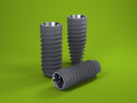
Atrophy of the mandibular bone caused by premature tooth loss due to periodontal or endodontic problems can often be found in posterior areas. Problems associated with implantation in these cases often arise due to limited bone height or width of the mandible, and several treatment options have been suggested to gain enough bone for a stable implantation and achieve an esthetically good result.1 In clinical practice, freeze-dried bone allograft (FDBA) blocks have been used for alveolar ridge augmentation with promising results, offering patients a less-invasive treatment alternative to autogenous bone blocks, with no donor site morbidity and no second surgical site.2,3,4 Nowadays, customized allogenic bone blocks can be produced using the computer-aided design/computer-aided manufacturing (CAD/CAM) technology, enabling a shorter surgery time as manual block adjustment during surgery becomes unnecessary, thus enhancing patient comfort.5,6 This case report describes a two-stage Guided Bone Regeneration (GBR) procedure using a customized allogenic bone block as a first step to increase the horizontal mandibular bone width. In a second step, special newly designed implants (Straumann® BLX Roxolid® SLActive®) were inserted to achieve good primary stability.
Initial situation
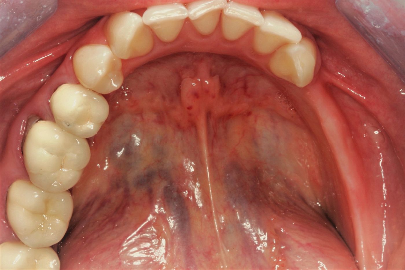
A 42-year-old woman presented with the wish for a fixed prosthetic rehabilitation in the lower jaw. The initial clinical and radiographic examination showed an atrophic jaw with limited bone availability for implantation (Figs. 1-2). Several treatment options for a two-stage GBR procedure to regain an optimal horizontal width for further implantation were discussed with the patient. In the end, because the patient refused autologous bone augmentation, treatment with a customized CAD/CAM freeze-dried bone allograft (maxgraft® bonebuilder, botiss biomaterials GmbH), followed by placement of Straumann® BLX Roxolid® SLActive® implants was determined.
Planning
A CBCT scan was taken and forwarded, in the Digital Imaging and Communications in Medicine (DICOM) data format, to botiss biomaterials GmbH to design the customized allogenic bone block.
botiss virtually designed the allogenic bone block on a three-dimensional reconstruction of the patient’s defect. After review of the block design and approval by the surgeon, the maxgraft® bonebuilder was milled from processed (Allotec® process, Cells + Tissuebank Austria (C+TBA), Krems, Austria) cancellous bone from femoral heads of living donors (Fig. 3).
Surgical procedure
The GBR procedure was performed under local anesthesia. A full-thickness vestibular flap with mesial relief incisions was raised. The lingual tissue was carefully dissected from the residual bone down to the mylohyoid muscle while protecting the neurovascular tissues without any sharp incision. So the lingual tissue was mobilized in the buccal direction for proper soft tissue management.
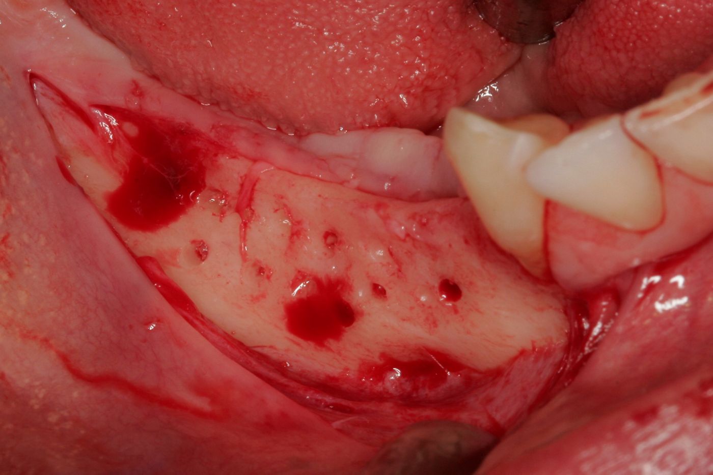
The cortical layer of the recipient site was perforated using a small round bur to promote bleeding and accelerate revascularization of the graft (Fig. 4). The customized allogenic block fitted exactly onto the recipient site and was rigidly fixed to the mandible with 1.25 mm wide and 8 mm long screws (Fig. 5).
Mesial and distal areas were contoured using xenogenic bone substitute material (cerabone®). The surgical area was covered with a pericardium collagen membrane (Jason® membrane), which was fixed to the local bone using titanium pins.
The flap was adapted and sutured using nonabsorbable 4.0 suture material. An apically positioned lateral mattress suture secured the muscle tension of the flap to achieve a tension-free wound closure. Sutures were removed at 14 days postoperatively.
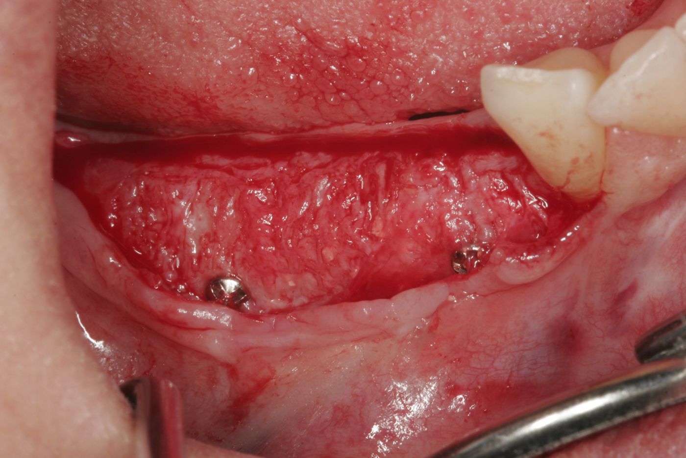
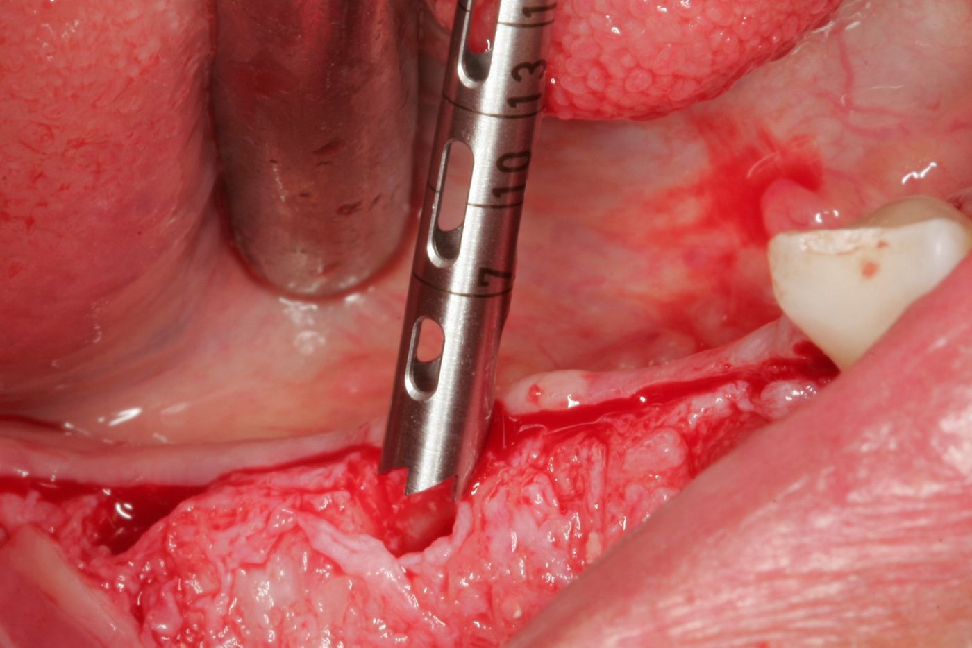
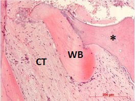
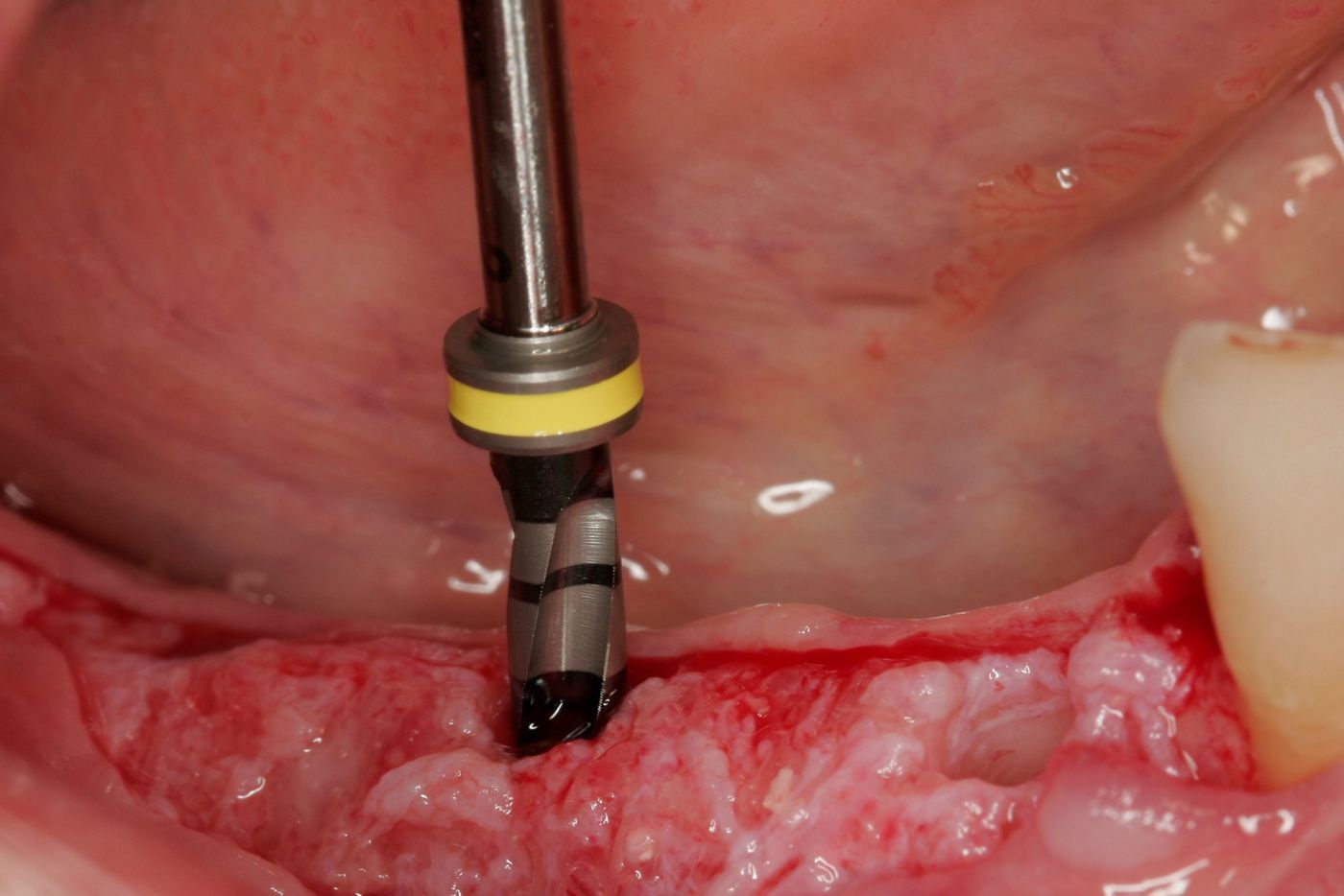
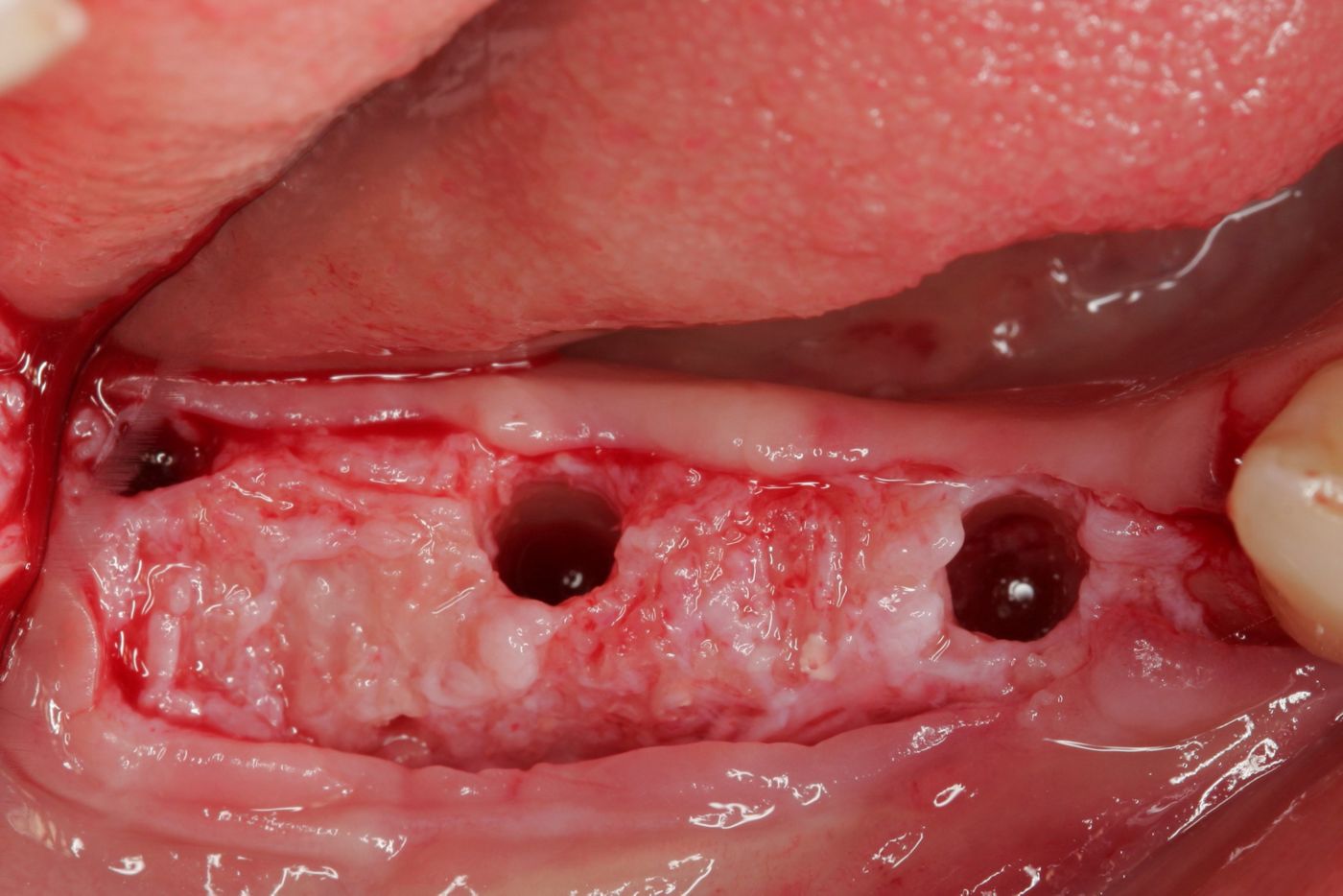
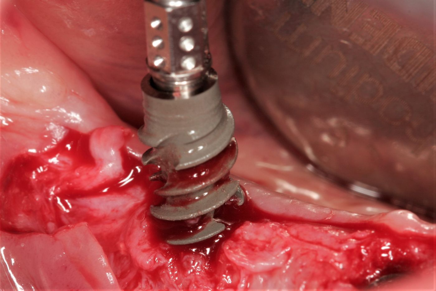
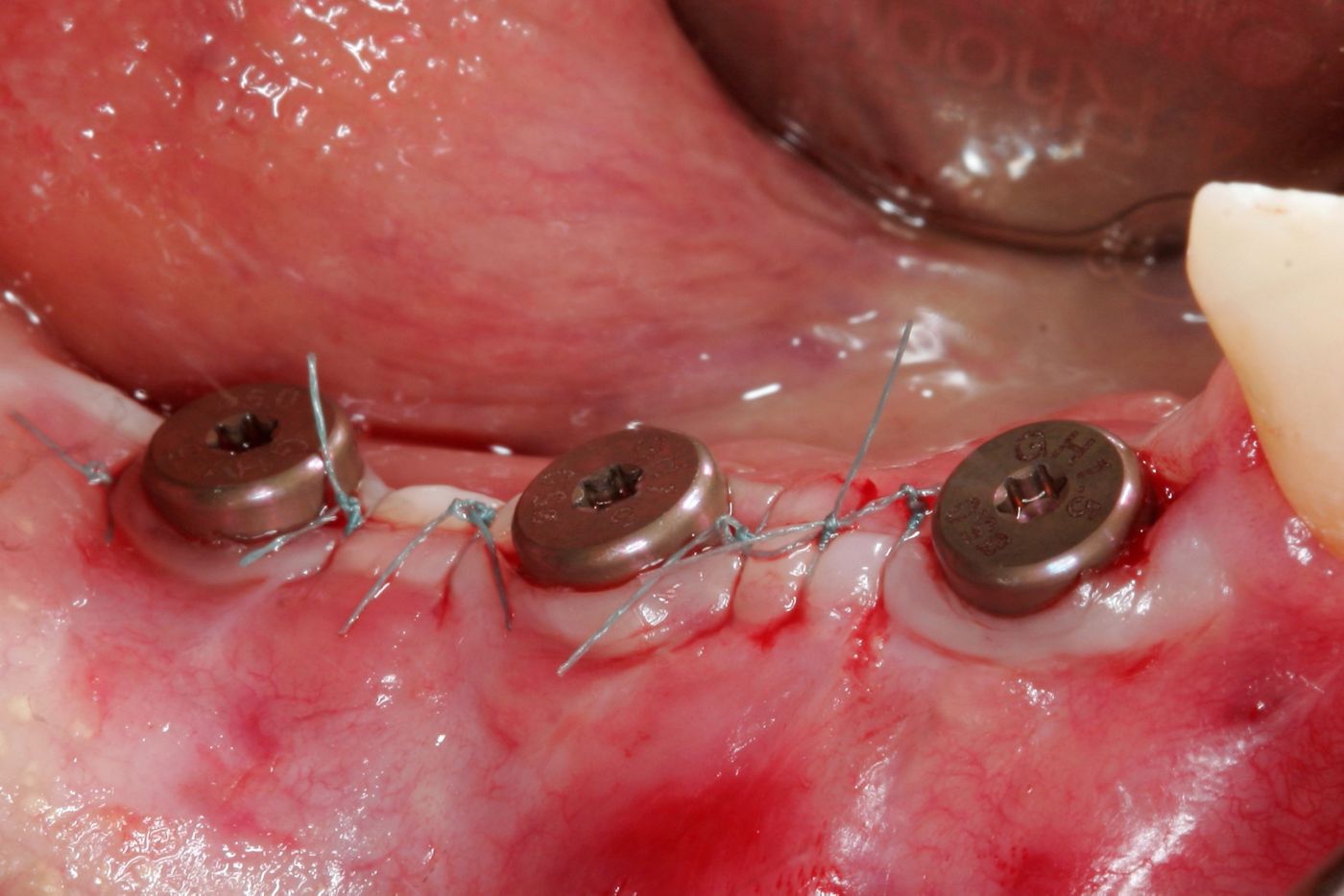
Following six months of uneventful recovery and healing, the patient presented for the implantation procedure (Figs. 6,7). At re-entry, the fixation screws were removed and a bone core biopsy was taken for histological analysis (Figs. 8-9). Biopsy slides were stained with hematoxylin and eosin stain, and the histological examination of the material obtained at re-entry showed the ongoing remodeling process of the FDBA block. Newly formed bone (woven bone, WB) was found to be in close contact with the allograft material (*) surrounded by connective tissue (CT), showing the material-mediated bone regeneration (Fig. 10) Three Straumann® BLX Roxolid®, SLActive® implants 4.5 mm in diameter and 10 mm long were inserted at bone level after measurements at locations 47, 46 and 44 (Figs. 11-14). Implants were covered with RB Closure Caps, and the surgical site was closed with 4/0 sutures (Figs. 15-16).
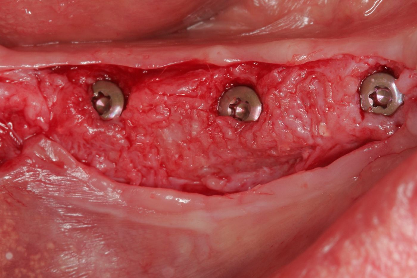
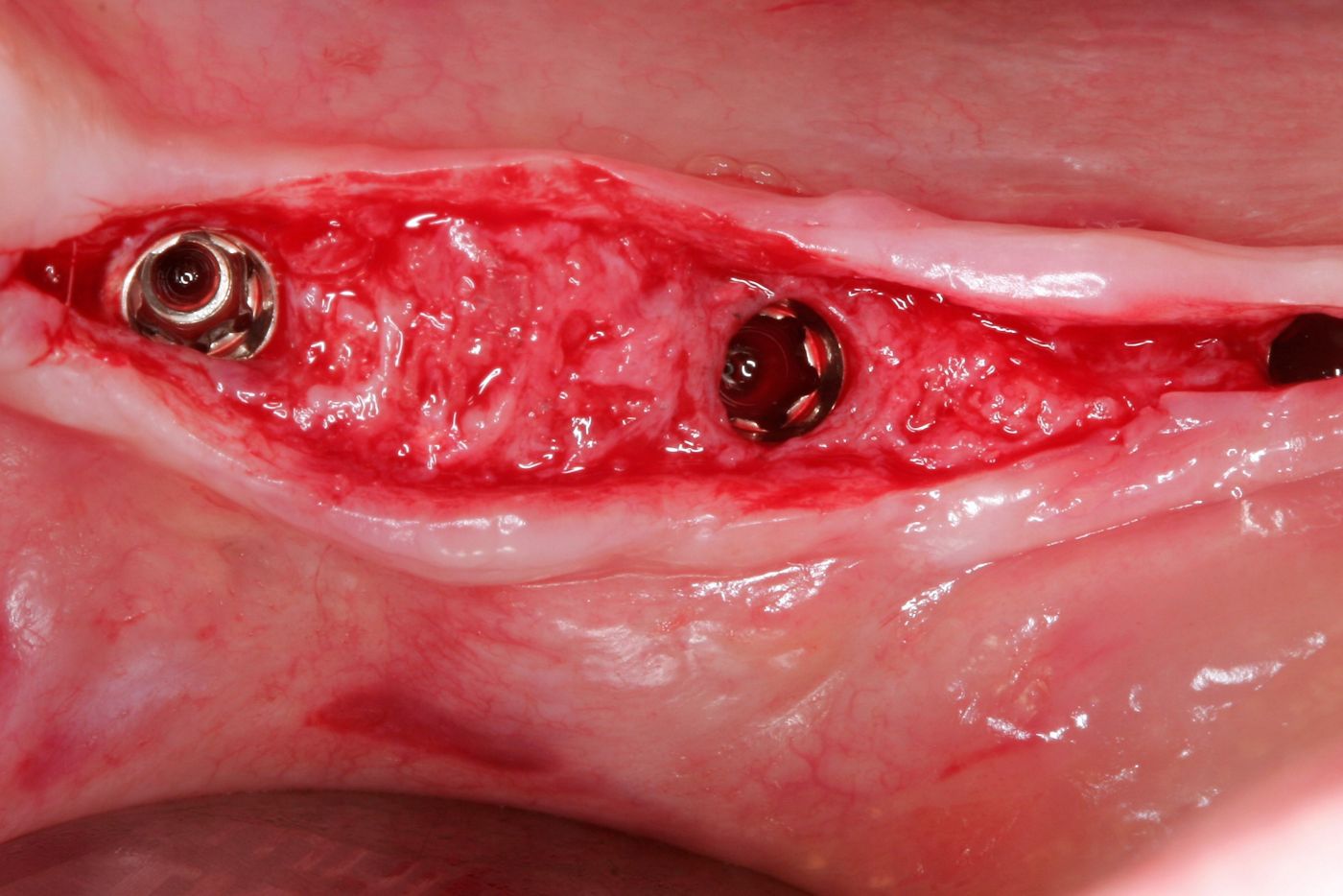
After three months, the implants were uncovered by a crestal incision. The Closure Caps were covered in places by new bone (Fig. 17). This shows the vital potential of the new generated bone in this area. RB/WB Healing Abutments were inserted (diameter 5 mm, 1.5 mm gingival height), and the soft tissues were approximated with 5/0 non-absorbable sutures (Figs. 18-20).
Prosthetic procedure
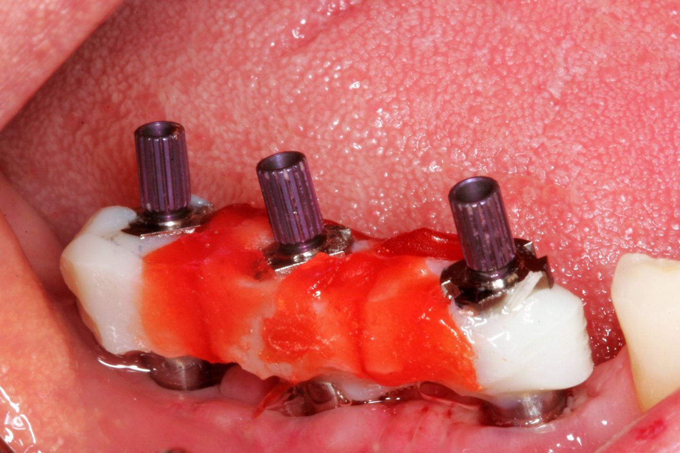
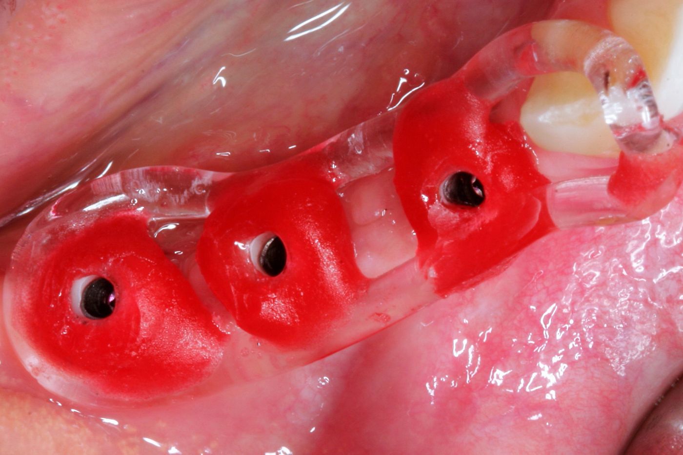
After three weeks of healing time, the impression was taken with splinted RB impression posts. An open-tray technique was performed in order to avoid dimensional changes during the transfer to the master model. An individualized open tray for the impression was used with Polyether Impression Material (Impregum Penta, 3M-ESPE) (Fig. 21).
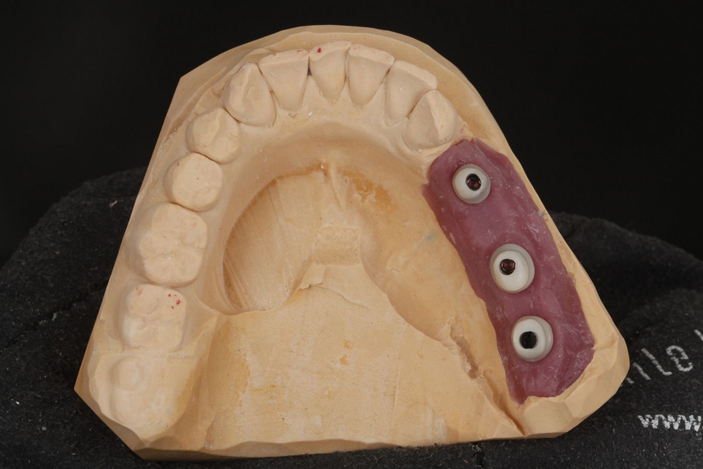
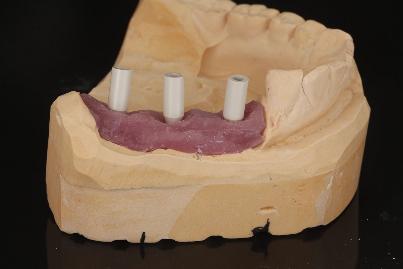
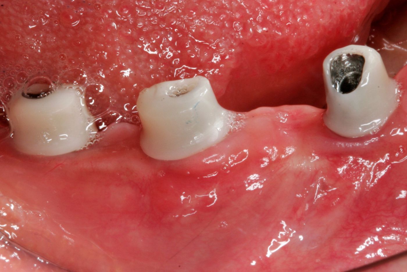
The individual abutments were created using a Variobase® Abutment with Zirconium Dioxide (Figs. 22-23). The Straumann® Variobase® Abutment provides dental laboratories with the flexibility to create customized abutments (Fig. 24). The preferred workflow was in-lab milling. The abutment combines the benefits of the original Straumann connection and the unique Straumann engaging mechanism.
An acrylic with pattern resin modified key was used to find the correct position of the abutments in the patient's mouth (Figs. 25-26).
Follow-up 10 months after implant placement showed a well- preserved gingival contour (Fig. 23-24).
Results
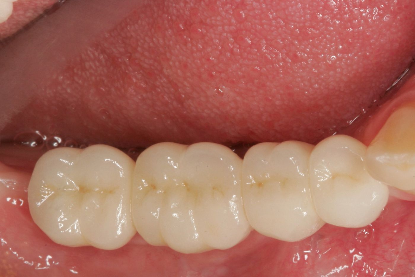
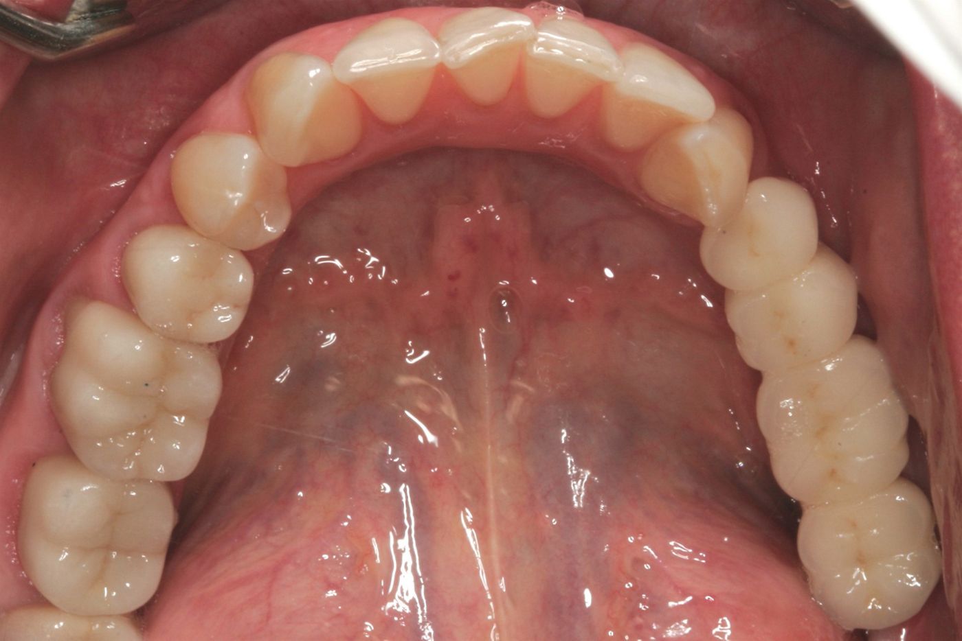
After successful integration of the prosthetic work, a zirconia and ceramic layered bridge was fixed with glass ionomer cement (Figs. 27-28).
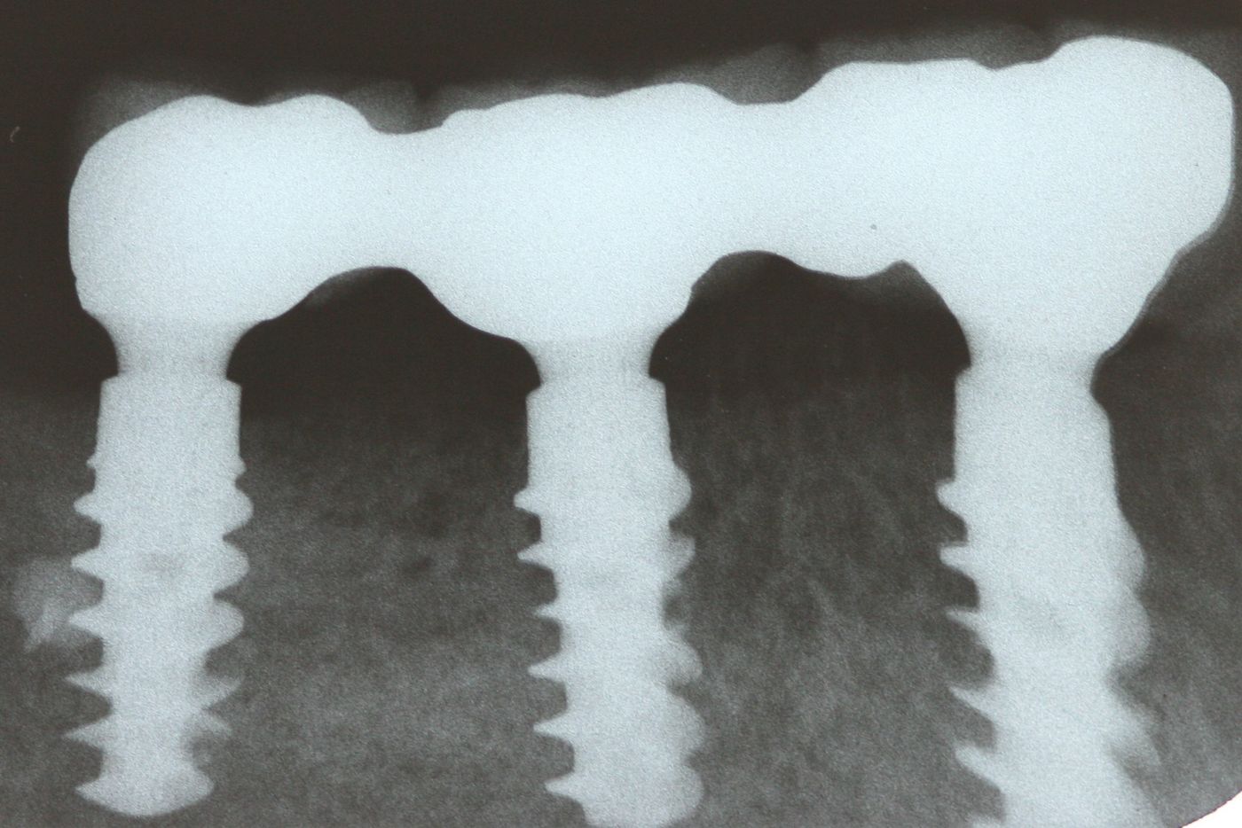
A check x-ray revealed a perfect fit of the prosthetic work (Fig. 29).
Conclusion
BLX implants achieved an optimal primary stability. BLX drills allow for adaptation of the implants’ primary stability by making intermittent movements during the bed preparation. From this perspective, this implant is very easy to use in different segments of the bone length and bone density characteristics, as in this case.
The two-stage GBR procedure using a customized FDBA block was able to fulfill the patient’s wish for fixed dental prostheses without the need for harvesting of autologous bone, saving the patient the need for additional surgical sites for bone harvesting. CAD/CAM technology produced an optimal fitting allogenic bone block, reducing the surgery time as no manual adjustment of the block was necessary, thus also reducing patient discomfort. The augmentation procedure gained significant horizontal bone width for successful implantation. With its special thread design, the BLX implant chosen here showed excellent cutting and fixation properties in such different bone situations over the length of the implant, where more dense residual bone meets newly built bone tissue. During uncovering, the newly formed bone tissue was even growing on top of the closure caps in places, showing the excellent remodeling properties of the allograft material.
Overall, this case demonstrates that, while being far less invasive, allogenic block augmentation, and especially the customized allogenic bone blocks, facilitate bone augmentation procedures for the surgeon and the patient. At the same time, these long-lasting implant solutions offer maximum comfort for the patient.
Straumann® BLX Implant System – new versatility and ease of use in immediate restorations
(Manufacturer information) To provide the most versatile and the easiest to use of all available implants for immediate protocols – this is what the new BLX system from Straumann is aiming for. All clinical experiences with fully tapered implants in the last decade, and of course with Straumann’s own apically tapered BLT implant, were taken into account during development. In order to combine in a single product high effectiveness in typical immediate restoration procedures with exceptionally forgiving characteristics in less than ideal situations, Straumann sought to achieve more than the design of a new fully tapered implant. The BLX concept relies on a combination of a set of unique and entirely new drills (Velodrill™) which prevents hyperthermia of the bone, as well as specific implant shapes that achieve high primary stability in soft bone, but without over-compressing the cortical areas. All BLX sizes have the same prosthetic connection, and the 3.75 mm diameter has already been cleared for all indications. The result is a next-generation implant system that aims to offer new levels of confidence—for immediate restorations and all other treatment protocols in order to suit the dentist´s preference. It also enables the use of smaller implants. Additionally, the resulting simplified workflows translate into shorter chair time. Having already attained very positive results and feedback, Straumann BLX combines a highly innovative design for primary stability with its high-performance Roxolid metal alloy and SLActive surface. Thus, the new BLX will bring both versatility and ease of use to immediate protocols.
