#Preventive 12. Nov 2024

Peri-implantitis: Effective electrolytic cleaning and regenerative therapy

A clinical case report by Algirdas Puišys, Lithuania

DDS and PhD from Vilnius University, – Periodontologist at the Lithuania University of Health Science. Engaged by the private practice VIC clinic in Lithuania. Course instructor and lecturer nationally and internationally. Fellow of the International Team of Implantology (ITI)

Introduction

Peri-implant diseases are inflammatory conditions that affect the soft and hard tissues surrounding dental implants. These conditions are categorized primarily into two types: peri-implant mucositis and peri-implantitis. Peri-implant mucositis is characterized by inflammation confined to the peri-implant soft tissues without bone loss. This condition is considered reversible with appropriate intervention. In contrast, peri-implantitis extends beyond the soft tissues, involving the progressive loss of supporting alveolar bone, which can severely compromise the structural integrity of the dental implant. Peri-implantitis therefore represents a significant threat to the long-term success and survival of dental implants and, if not managed effectively, can ultimately lead to implant failure and loss.1,2

The pathogenesis of peri-implantitis is closely linked to the formation and maturation of a bacterial biofilm on the implant surface. This biofilm, a complex and resilient community of microorganisms encased in an extracellular polymeric matrix, is a critical etiological factor in the onset and progression of peri-implant diseases.3 The host immune response to these biofilms is characterized by the stimulation of inflammatory cells, including neutrophils and macrophages, which release pro-inflammatory cytokines and enzymes that degrade bone tissue. This inflammatory cascade leads to peri-implant bone resorption, jeopardizing the stability of the implant.4 Given the central role of biofilm in disease progression, effective biofilm removal is key in the management of peri-implantitis.

Treatment strategies for peri-implant diseases have evolved to include various approaches, often requiring a combination of mechanical debridement, antimicrobial therapy, and, in more advanced cases, surgical intervention. Mechanical debridement remains a cornerstone of treatment, focusing on the physical removal of biofilm and calculus from the implant surface. This may involve the use of specialized instruments such as ultrasonic scalers, titanium curettes, or air-polishing devices. In recent years, electrolytic cleaning has emerged as a novel approach for biofilm removal, utilizing low-level electrical currents to disrupt and detach bacterial biofilms from the implant surface.

However, in cases of established peri-implantitis, non-surgical approaches may be insufficient to fully resolve the disease, and surgical interventions are often necessary to achieve thorough decontamination of the implant surface and to facilitate the regeneration of lost peri-implant tissues. The goal of these procedures is to re-establish a stable and healthy peri-implant environment conducive to the long-term stability of the implant. 5,6

In this clinical case, we describe the successful management of peri-implantitis through a comprehensive treatment that integrates meticulous mechanical cleaning with regenerative techniques. This case highlights the critical importance of the efficacy of biofilm removal strategies and the application of regenerative procedures to achieve optimal clinical outcomes. The integration of these techniques not only stops the progression of the disease, but also facilitates the regeneration of lost bone and soft tissue, thereby ensuring the long-term stability and function of the dental implant.

Initial situation

A 40-year-old female, healthy (ASA I), non-smoker, taking no medication, came to our practice in 2020 from a referral dentist and with a diagnosis of peri-implantitis in the posterior area. The patient mentioned that the implants were placed 7 years ago, and that she failed to attend her follow-up appointments. The patient requested to keep her implants.

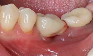
On intraoral examination, the implants in positions #36 and #37 showed a probing depth greater than 6 mm, positive BOP (Bleeding on Probing), suppuration, redness and swelling, and dental plaque.

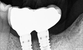
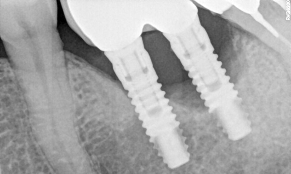
The radiographic examination revealed both horizontal and vertical bone loss around the implants, particularly around implant #37 (Fig. 1).


Fig. 1

Treatment planning

The treatment started with a non-surgical approach to reduce inflammation. Next, a surgical procedure was performed to decontaminate the implants, followed by guided bone regeneration (GBR). A new screw-retained splinted crown on the implants was then placed.

The treatment workflow included:

  1. Non-surgical periodontal supportive therapy: oral hygiene instructions, rinsing with 0.12% chlorhexidine (CHX), and the application of metronidazole at a concentration of 5 mg/ml, along with the use of a solution of local antibiotic and hyaluronic acid. Follow-up appointments were scheduled and conducted on the following dates: November 11, 2020; December 9, 2020; January 6, 2021; February 2, 2021; March 3, 2021. 
  2. Surgical treatment, which involved GalvoSurge®, GBR using autogenous bone chips and Straumann® Membrane Flex.
  3. Final prosthetic rehabilitation with screw-retained splinted crowns on implants.
  4. Follow-up visits for control


Surgical procedure

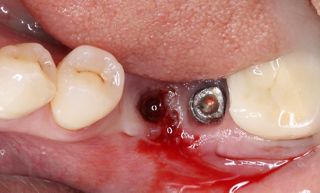
After the non-surgical treatment, clinical examination in 2021 showed a decrease in inflammation, though it was not completely resolved (Fig. 2). Lidocaine 2% with epinephrine 1:100,000 was administered. Upon removal of the prosthesis, the peri-implant mucosa surrounding the implants in positions #36 and #37 exhibited localized inflammation with evident redness, swelling and bleeding (Fig. 3).


Fig. 2

Fig. 3

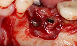
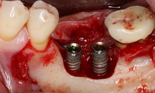
A full-thickness flap was elevated to access the defect and mechanically remove the granulation tissue surrounding the implants (Fig. 4). The bone defect was assessed using the modified criteria established by Monje et al, and was classified as a Class 3b defect, making it suitable for reconstructive therapy. First, implant disinfection was done with ablative mechanical debridement, CHX 0.12 %, metronidazole 5mg/ml, and a solution of local antibiotic and hyaluronic acid (Fig. 5).


Fig. 4

Fig. 5

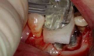
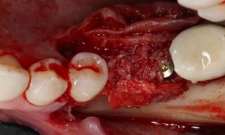
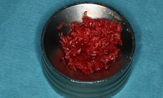
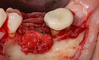
The implant cleaning and disinfection were done with CHX 0.12 % and the GalvoSurge®. Implant disinfection was performed using GalvoSurge®, applying only gentle pressure to the implant being treated. In this 2-minute process, hydrogen ions interact with captured electrons to produce hydrogen bubbles that lift the biofilm off the implant surface (Fig. 6). Additionally, regenerative therapy was performed through GBR. Autogenous bone chips, combined with bone harvested from the tuber, were placed into the defect as grafts. These materials helped in bone augmentation and covered the exposed implant threads, thereby enhancing the healing and re-osseointegration at the implant site (Figs. 7-9).


Fig. 6

Fig. 8

Fig. 7

Fig. 9

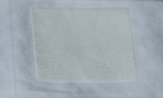
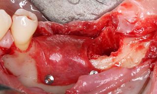
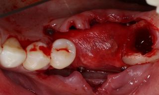
Tooth #38 was extracted. Closure caps were placed, and the Straumann® Membrane Flex, a minimally-crosslinked porcine peritoneum collagen membrane, was fixed in place with pins. This protects the graft area from unwanted soft tissue infiltration during the healing phase (Figs. 10-12).


Fig. 10

Fig. 12

Fig. 11

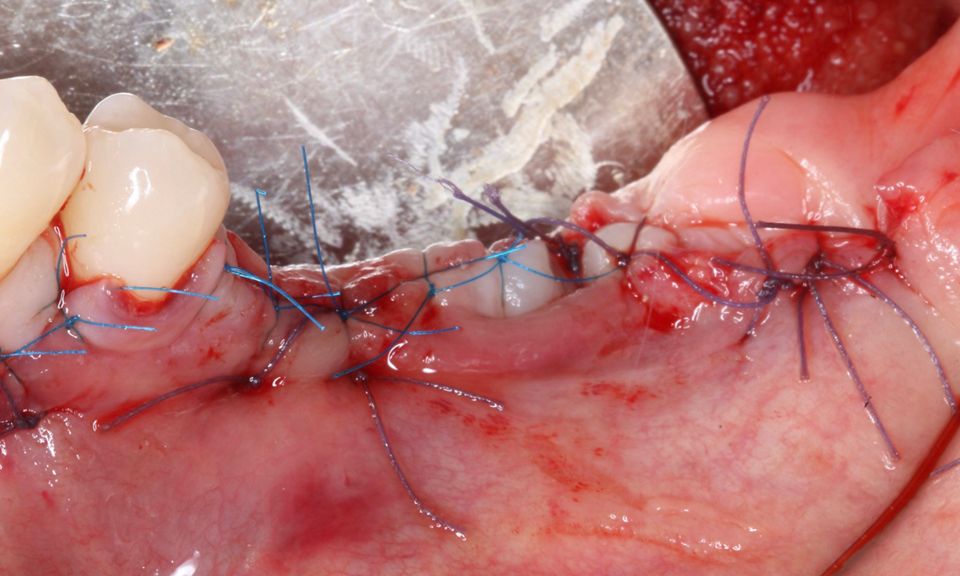
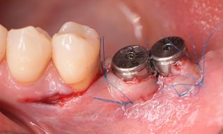
Suturing was performed with 4/0 vicryl and 6/0 prolene, using interrupted sutures to facilitate primary intention wound healing, while the implant remained without the prosthetic crown for 6 months (Fig. 13). After this period of submerged healing, the reinstallation of the suprastructures was planned.


Fig. 13

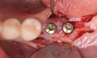
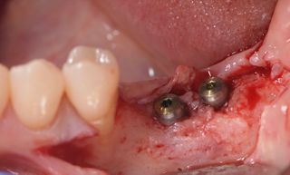
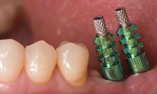
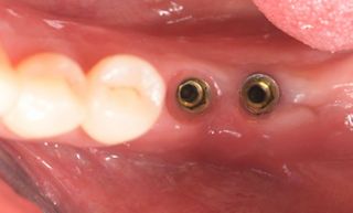
In 2021, six months after the initial surgery, a full-thickness flap was elevated, revealing that the implants were surrounded by bone (Figs. 14,15). Healing abutments were then inserted, and interrupted sutures were inserted using 6/0 prolene (Fig. 16). The healing abutments play an essential role in facilitating proper healing of the gingival tissue around the implant, shaping the tissue for an optimal fit of the final prosthesis, and protecting the implant from contaminants.


Fig. 14

Fig. 16

Fig. 15

Prosthetic procedure

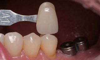
After the healing period, the next step involved selection of the appropriate shade for the fully polished zirconia prosthetic to ensure esthetic integration with the surrounding natural dentition. To achieve a precise match and optimal visual outcome, a shade guide was used, and the selected shade was confirmed with the patient (Fig. 17). An impression was taken with the open-tray technique, using addition silicone material for its high dimensional stability and accuracy (Fig. 18). Then it was sent to the dental laboratory for the fabrication of the final prosthesis.


Fig. 17

Fig. 18

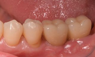
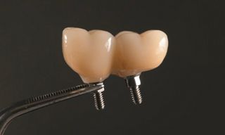
The gingiva was evaluated before the placement of the final prosthetics (Fig. 19). A fully polished zirconia prosthetic was then carefully fabricated and inserted, and torqued to 35 Ncm (Figs. 20,21) and the subsequent x-ray revealed satisfactory results (Fig. 22).


Fig. 19

Fig. 21

Fig. 20

Fig. 22

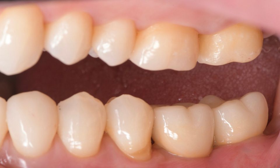
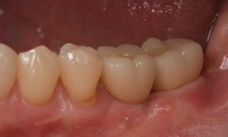
At the follow-up recall in 2021, the clinical evaluation indicated that the peri-implant soft tissues were in good health, with no signs of inflammation. Additionally, the marginal levels were appropriately maintained (Fig. 23).


Fig. 23

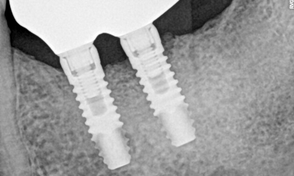
The following year, the clinical evaluation revealed similar findings, with the peri-implant soft tissues remaining healthy. The radiographic examination also showed that the peri-implant marginal bone levels were stable, with no evidence of bone resorption (Fig. 24).


Fig. 24

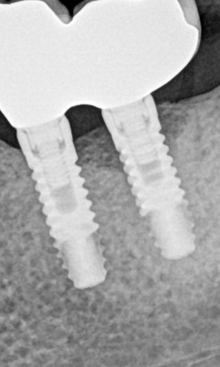
In 2023, similar results were observed, with the peri-implant tissues remaining healthy. The radiographic examination confirmed that marginal bone levels were intact. The treatment was successful, and the patient reported satisfaction with the esthetic and functional outcomes. (Figs. 25,26).


Fig. 25

Fig. 26

Treatment outcomes

The follow-up visits confirmed the absence of any biological or radiographic issues, demonstrating excellent health in both hard and soft tissues. This outcome highlights the success of the surgical procedure, further improved by the use of GalvoSurge® and GBR.

Author’s testimonial

I think the GalvoSurge® device is a great tool for disinfecting the implant surface, which can help re-establish osseointegration. However, it's important not to forget the importance of conservative treatments as a first step and regular follow-up appointments.

References:

  1. Schwarz, F., et al. "Peri-implantitis." Journal of Clinical Periodontology, vol. 45, no. 20, 2018, pp. S246-S266.
  2. Lang, N. P., et al. "Peri-implant diseases: where are we now?–Consensus of the Seventh European Workshop on Periodontology." Journal of Clinical Periodontology, vol. 38, no. S11, 2011, pp. 178-181.
  3. Renvert, S., and N. Polyzois. "Risk indicators for peri‐implantitis. A narrative review." Clinical Oral Implants Research, vol. 26, no. 11, 2015, pp. 15-44.
  4. Koyanagi, T., et al. "Immunological and microbiological aspects of peri-implant diseases." Journal of Clinical Periodontology, vol. 40, no. 9, 2013, pp. 887-895.
  5. Heitz-Mayfield, L. J. A., and T. Mombelli. "The therapy of peri-implantitis: a systematic review." International Journal of Oral and Maxillofacial Implants, vol. 29, 2014, pp. 325-345.
  6. Smeets, R., et al. "Definitions, etiology, prevention and treatment of peri-implantitis–a review." Head & Face Medicine, vol. 10, no. 1, 2014, pp. 1-13.