

Immediate implant placement offers notable benefits, such as a reduced number of surgical interventions, a shorter overall treatment duration, and improve esthetic outcomes. This approach also preserves the existing bone and gingival structure, contributing to the support of interdental papillae.2
However, reaching and maintaining optimal gingival esthetics around implants in the anterior region is a challenging task. Ensuring sufficient primary stability is a prerequisite for the success of this approach. The design of the implant itself is a crucial factor. Recently, the findings from a series of cases indicated that the immediate placement of a novel self-cutting, tapered implant (Straumann® BLX, Institut Straumann AG, Basel, Switzerland) with immediate provisionalization through an integrated digital workflow, can yield reliable functional and esthetic outcomes when transitioning compromised single teeth in the esthetic zone.3
The Straumann® BLX Implants are made from Roxolid® material with the SLActive® surface. The use of Roxolid® material allows for the placement of reduced-diameter implants while ensuring successful osseointegration. Moreover, the integration of SLActive® surface technology accelerates osseointegration and minimizes the healing period.
The following case report outlines a successful treatment result for a compromised tooth in the esthetic region, characterized by a thin gingival biotype. The treatment involved the utilization of the Straumann® BLX Implant System, along with botiss cerabone® and botiss mucoderm®, with a digital workflow.
Initial situation
A young and healthy 25-year-old male patient, a non-smoker, presented at our clinic due to the fracture of his crown on the upper right lateral incisor. The patient was seeking a prompt, durable, and esthetic solution.
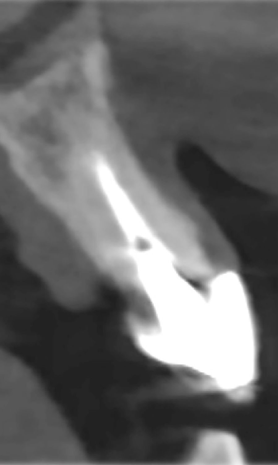
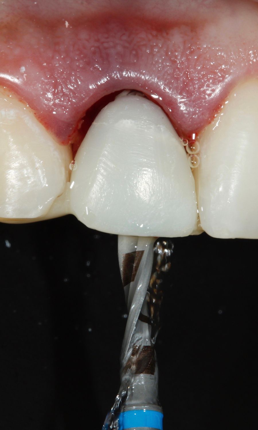
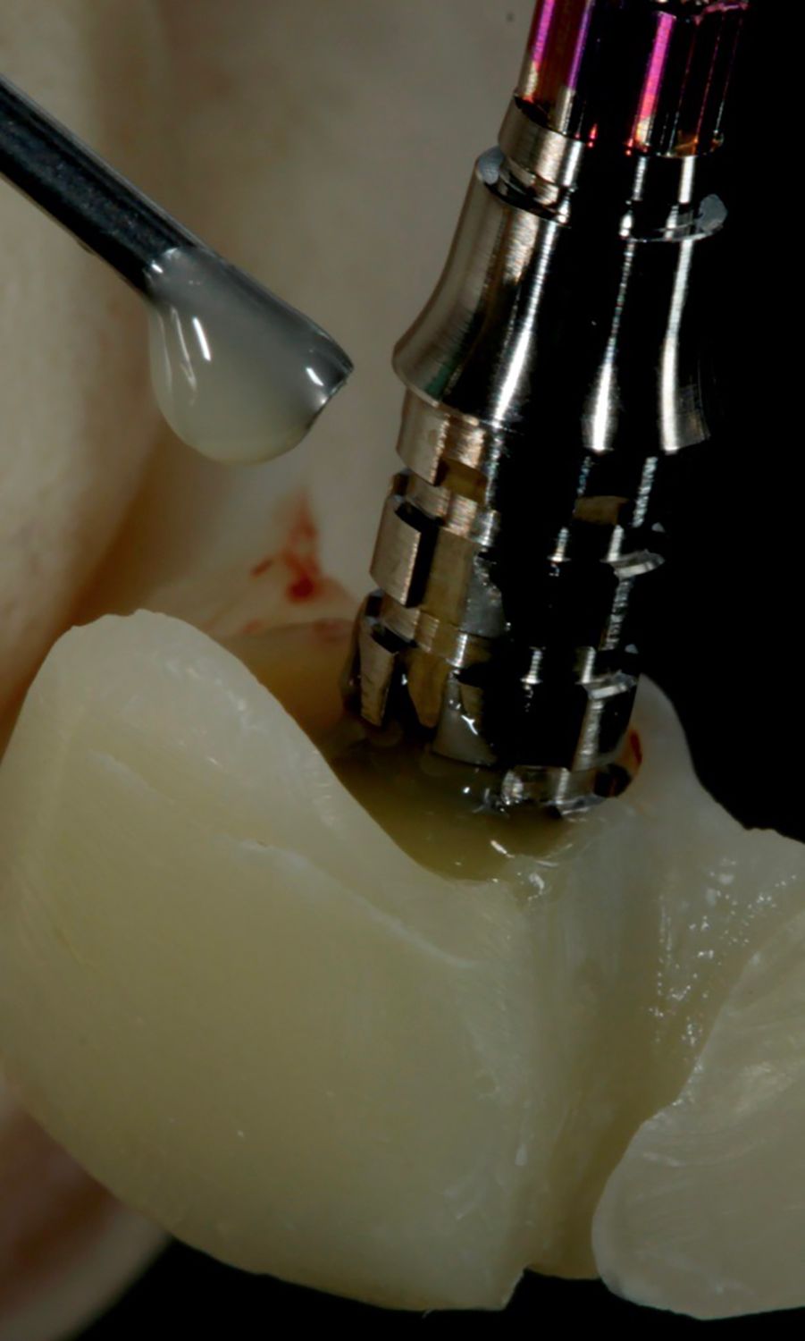
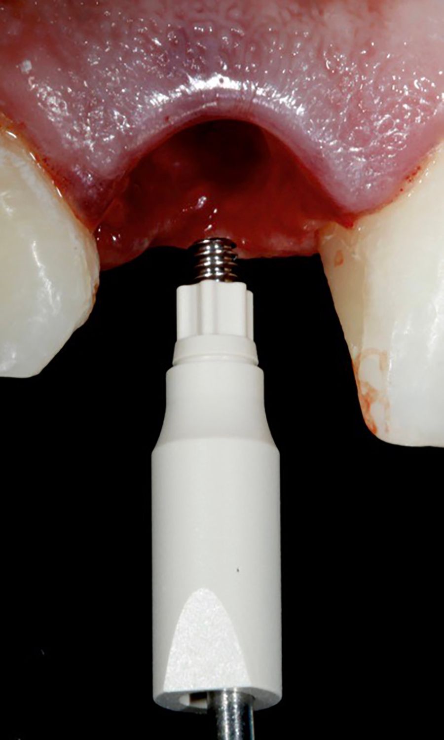
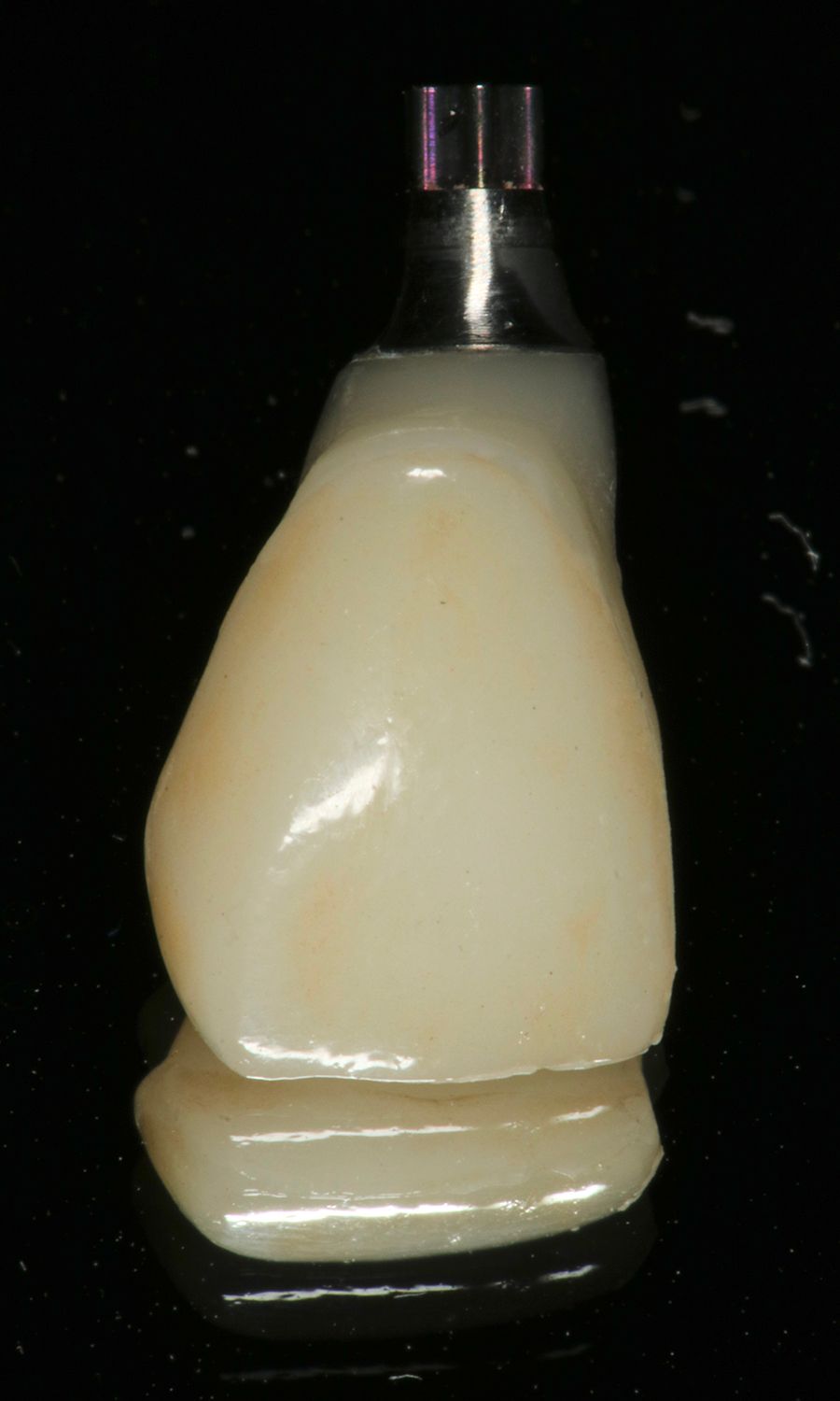
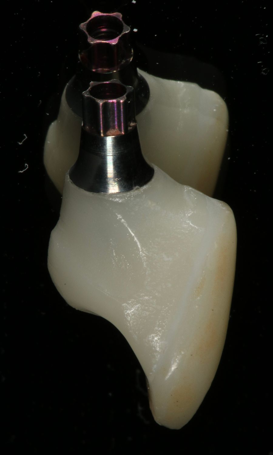
The extraoral examination showed a medium smile line. On intraoral examination, a metal-ceramic crown with chipping on the palatal side was observed on tooth #12 (Figs. 1,2).


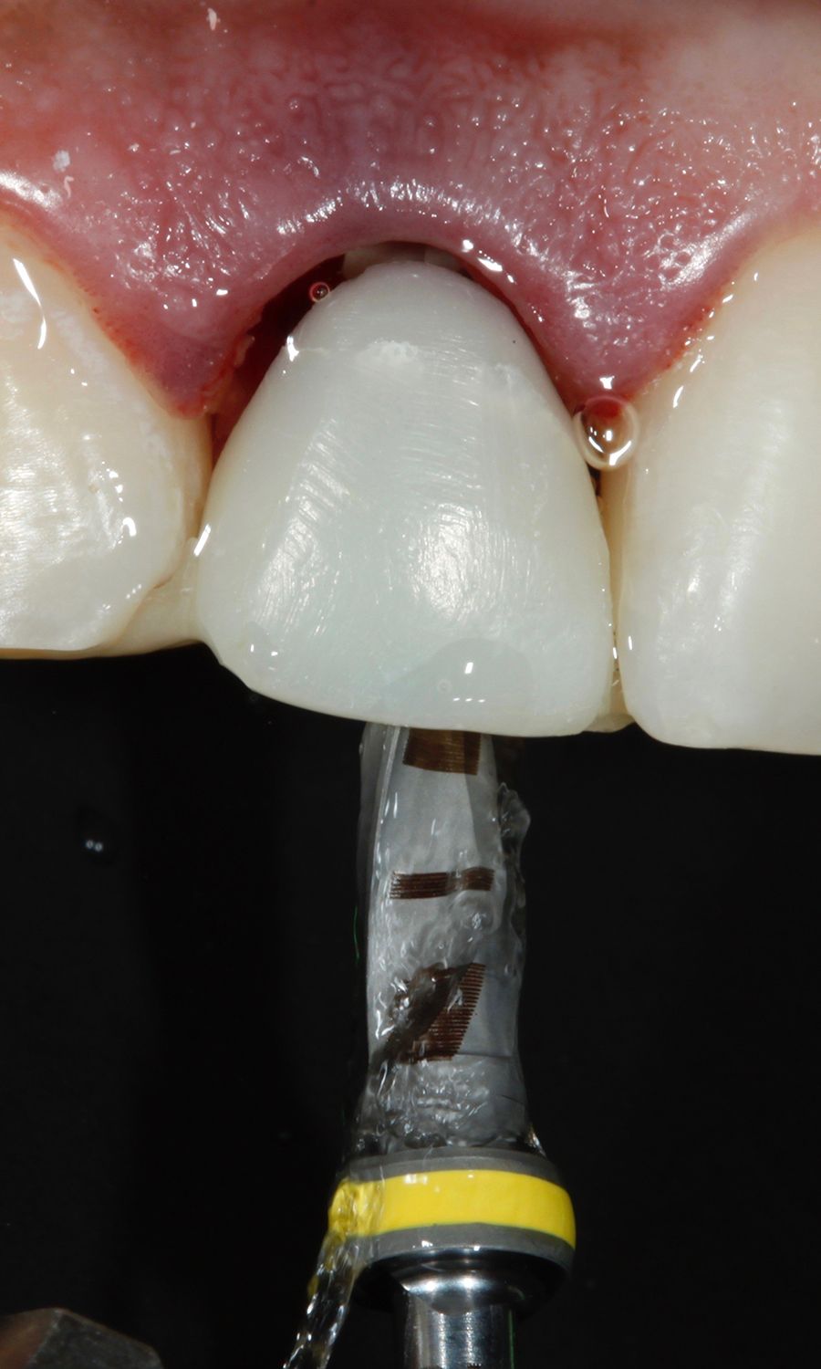
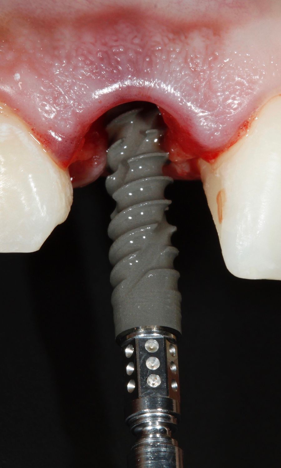
After the crown was removed, there was not enough stump left. The tooth was listed as hopeless. Additionally, signs of gingival inflammation around the residual root were noted (Figs. 3,4).