A 58-year-old healthy female patient presented to our office with the chief complaint of reduced masticatory function on the left side and the desire to improve her smile. Moreover, the patient expressed her wish to avoid removable rehabilitations during any of the treatment phases and wanted to be treated with a minimally invasive procedure, as she has had previous unpleasant experiences with dental treatments. The extraoral evaluation revealed a medium smile line with a dark area on the second quadrant due to the absence of premolars and molars (Fig. 1).

The intraoral assessment showed a partially edentulous maxilla, with missing teeth #17, #14, #24, #25, #26 and #27 and black triangles on the second sextant (Fig. 2). The occlusal view of the upper jaw showed a Kennedy Class II Applegate modification I with slightly misaligned teeth (Fig. 3). The lateral photo showed a possible good amount of available bone in terms of height (Fig. 4). The view of the left side of the mouth in occlusion showed the extrusion of teeth #34 and #36 (Fig. 5).


A CBCT scan visualized with the software coDiagnostiX® was recorded to assess the quality and quantity of bone available for implant placement (Fig. 6). In the region #24-#26, the assessment revealed adequate vertical and horizontal bone availability.

Treatment planning Considering the patient’s requests, the aims of the treatment were:
- To evaluate the placement of two implants in positions #24 and #26 to be restored with a fixed three-unit bridge.
- To consider the possibility of placing an immediate prosthesis on the implants, if satisfactory primary stability is obtained: in this way, a pleasant esthetic appearance is guaranteed from the beginning of the treatment.
- To consider the option of placing the implants with computer-guided surgery and a flapless approach to reduce the invasiveness and to simplify the prosthetic workflow.
Taking the above aims into consideration, the following treatment options were presented and discussed with the patient:
- Implant placement in positions #24 and #26 with no provisional restoration during the healing time. After implant osseointegration, final restoration with a three-unit implant-supported bridge #24-#26.
- Implant placement in positions #24 and #26 and, if the implant stability is optimal, an immediate provisional implant-supported bridge. After implant osseointegration, final restoration with a three-unit implant-supported bridge #24-#26.
- Implant placement in positions #24 and #26 with a guided surgery approach and, if the implant stability is adequate, an immediate provisional implant-supported bridge. After implant osseointegration, final restoration with a three-unit implant-supported bridge #24-#26.
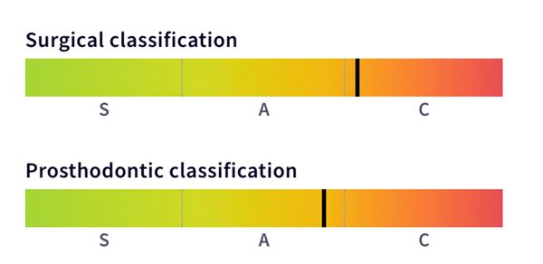
The SAC v2.0 Assessment Tool (based on ITI SAC Classification) was used to identify the degree of complexity and the potential risk involved in the planned case (Fig. 7).
In order to make the final decision on treatment planning, the pros and cons were evaluated taking into account the choice of computer-guided implant positioning with an immediate prosthesis (three-unit bridge) for the replacement of teeth #24, #25 and #26.
It was decided to choose option number 3 after discussing the risks, benefits, and treatment options, and considering the patient's age, medical conditions, and expectations. Thus, it was planned to treat the patient with Straumann® BLX implants in sites #24 and #26 with immediate prostheses (three-unit bridge) using computer-guided planning and surgery. The rationale for the treatment was as follows:
- The choice of a fixed implant-supported rehabilitation satisfied the patient’s request to avoid a removable prosthetic solution.
- The patient can be provided, as requested, with a fixed provisional bridge assuring a pleasing esthetic appearance from the beginning of the treatment.
- Thanks to their specific shape, the use of BLX implants guarantees the high stability needed for the immediate prosthesis.
- The computer-guided planning and implant placement assure a surgical approach with reduced invasiveness and simplify the prosthetic procedures.
- The appropriate amount of keratinized gingiva allows for a flapless approach, thus minimizing the surgical impact.
- Favorable site anatomy, suitable bone availability, and a convenient amount of keratinized gingiva reduce the risk of esthetic complications.
Treatment workflow included:
- Preliminary data acquisition: Intra- and extraoral photos, impressions, creation of the diagnostic guide, and CBCT scan with the diagnostic guide in position (already collected).
- Scanning of the cast model and the diagnostic guide seated on the model; creation of the corresponding STL files and the STL file related to the digital wax-up of the missing teeth.
- Processing of Dicom (CBCT scan) and STL (upper model, upper model with guide and digital wax-up) data in coDiagnostiX® planning software to carefully plan the implant placement.
- Production of printed surgical guide and resin models via the coDiagnostiX® plan.
- Fabrication of the provisional bridge by the dental lab based on digital wax-up (on resin printed models).
- Surgical phase: implant placement and provisional bridge positioning.
- After the healing, final rehabilitation with a screw-retained bridge on the implants.
Surgical procedure
The patient was instructed to rinse her mouth with 0.12% chlorhexidine gluconate on the day of surgery. Anesthetic infiltration was done with 2% lidocaine and 1:100,000 epinephrine in the area corresponding to the premolar/ molar apexes and in the surrounding gingiva (Fig. 8).

A dedicated set of surgical instruments for the BLX implant guided surgery was used (Figs. 9,10).


The guide was placed in the mouth, and the stability and precision were verified through the windows created in the guide at specific teeth positions (Fig. 11). The lateral view of the guide shows the sleeve dedicated to the pin fixation, which was essential to guarantee the perfect stability of the guide during the drilling procedures (Fig. 12). From the occlusal view of the guide, the areas of keratinized gingiva visible through the sleeves assured the possibility of performing flapless implant surgery (Fig. 13).


A tissue punch, directed by the guide, was used with the purpose of removing the soft tissue layer. During this phase there was no need to fix the guide with the fixation pin (Fig. 14). Then the guide was removed, and it was easy to see the circular cut of the tissue punch (Fig. 15). The soft tissue disks were then peeled away (Fig. 16).


The guide was kept in its precise position and, using the dedicated bur, the drilling procedure through the sleeve was then performed (Figs. 17,18).


The fixation pin, placed in the dedicated sleeve, allows the guide to remain in its stable position, and the drilling procedure can be carried out in a precise way. After the guide placement (Fig. 19), the milling cutter bur was used with the purpose of creating a flat surface for the precise work of the following drills. The specific handle reduced the dimension of the sleeve to that of the selected drill. This bur is the only one without a stop (Fig. 20).


The ø 2.2 mm pilot drill was used (Fig. 21), followed by the ø 2.8 mm drill (Fig. 22), and finally the ø 3.5 mm drill (Fig. 23). Each drill was cooled with copious amounts of sterile water, and the tip was moved back and forth to minimize overheating.


In accordance with the choices made during coDiagnostiX® planning, a BLX implant 4.5 mm in diameter and 12 mm long was used in position #24 (Fig. 24). The pin placed in the apical part of the implant was gently broken according to the manufacturer’s instructions (Fig. 25).


The implant was then ready to be engaged into the sleeve for the perfect guidance in the prepared bone site (Fig. 26). The dedicated BLX transfer piece drives the implant in the correct position. The procedure ends as soon as the black line is in contact with the edge of the sleeve (Fig. 27).


The molar site was prepared following the procedure for the premolar. Because the quality of the bone was not excellent in this site, the ø 2.8 mm drill was the last one used. As seen in the procedure for the premolar, the implant was chosen in accordance with the coDiagnostiX® planning (BLX 4.5 mm in diameter and 8 mm long).
After placing the implant, the insertion torque obtained was evaluated. The high values reached allowed us to proceed with the immediate provisional bridge (Figs. 28,29).


In the occlusal view of the implants after placement, we observed an optimal 3D position (Fig. 30). The temporary abutments, customized by our dental lab, were screwed onto of the implants (Fig. 31). The temporary bridge was then checked in the patient's mouth in order to evaluate its precise seating on top of the temporary abutments (Fig. 32). The seating of the temporary bridge was very precise thanks to the accurate planning and execution of implant placement, perfectly in line with the prosthetic design (Fig. 33).


Prior to connecting the bridge and the abutments with resin, the field was isolated by means of two pieces of dental dam. This procedure is very useful especially when a flap is raised, and it is not easy to obtain the ideal wet conditions for resin polymerization (Fig. 34). Next, a thin layer of resin was applied to the temporary bridge (Fig. 35).


We can observe the temporary bridge in position, after the removal of the resin sheet that has allowed the precise placement of the bridge (Fig. 36). After the temporary bridge was unscrewed from the mouth, it was filled with resin in the spaces between bridge and abutments, and this was followed by the refinement procedures (Fig. 37).


Want to stay up to date?
youTooth.com is THE PLACE TO BE IN DENTISTRY – subscribe now and receive our monthly newsletter on top hot topics from the world of modern dentistry.
The refinement procedure for the temporary bridge was performed first with the tungsten carbide bur, then with a rubber pre-polisher and, finally, with a polishing buff. It is important to create an ideal emergence profile of the bridge units and to have a completely smooth surface in contact with soft tissues (Fig. 38). A reverse angle view of the temporary bridge showed the correctness of the emergence profiles: in this way the bridge started the precise conditioning of soft tissues from the beginning of the implant healing (Fig. 39).


The temporary bridge was screwed onto the implants (Figs. 40,41).


The chimneys were closed, and the smile line looked harmonious with the prosthesis well integrated into the patient’s mouth (Fig. 42). The temporary bridge was intentionally not in full occlusal contact in order to reduce the risk of excessive loading on implants (Fig. 43).


The patient’s smile immediately after the surgery (Fig. 44).

The treatment at this point already met the patient's expectations, delivering the expected esthetic and functional clinical benefits.
Prosthetic procedures
The patient reported no mechanical or biological issues at the 3-month follow-up visit. Moreover, the clinical examination showed that the emergence profiles created by the provisional bridge were harmonious and natural. To prepare the final restoration, the temporary bridge was removed, and the soft tissues were evaluated. The tissues around the implants were healthy and with an optimal emergence profile (Fig. 45). Afterwards, a conventional impression was taken (Fig. 46).


The final restorations were screwed onto implants 24 and 26, and the occlusal plane was corrected by making new restorations in the opposite jaw (Figs. 47,48). The final x-ray image showed the perfect integration of the implants and the precision of the prosthetic work (Fig. 49).


Treatment outcomes
The treatment outcome met the patient's esthetic and functional expectations. The patient reported an improvement in her quality of life. She was involved in a maintenance program with yearly follow-up visits. Two years later the patient is still very satisfied with the treatment outcome, and the peri-implant tissues appear in excellent condition (Figs. 50,51)


Acknowledgements:
I would like to express my gratitude to Dr. Elisa Oneto for her great contribution to the prosthetic procedures and to Alessandro Giacometti for his excellent lab work.
Dr Piano’s Testimonial
When indicated, immediate treatments can reduce the chair time and cost, maintain the gingival tissues and increase the comfort of our patients.