#Immediacy 01. Sep 2022

Soft tissue cantilever management in the aesthetic zone, replacing central and lateral upper incisors with a provisional prosthesis

A clinical case report by Alejandro Treviño Santos, México. Consuelo Montes Urrutia, Chile.

One of the major challenges in esthetic dental therapy is soft tissue management. The use of one implant with cantilever instead of two implants, seems to improve the formation of a more natural gingival contour and the preservation of inter-proximal papillae [1]. Considering that in between two implants, alveolar ridge is lost and therefore papillae conformation turns more complex [2]. An immediate interim prothesis —which is achievable thanks to immediacy provided by Straumann® BLX— allows correct soft tissue conformation. Even though cantilever generates lever force increased 32 to 64 times, this can be compensated with an anterior group/canine guidance. Specifically for anterior superior teeth, cantilever using group guidance, is an excellent choice to consider, without compromising implant stability and reaching an auspicious long-term prognosis [3]. Having a hybrid cemented-retained and screw-retained prosthesis, allow excellent and passive fit, lowers stress, and prevents from cement excess [4,5].

Initial situation

A healthy 27-year-old male suffered the avulsion of both central and lateral upper right incisors, during a street attack. At first, he attended to an emergency clinic in which the remaining teeth were splinted, and no effort was made to replace the avulsed teeth. (Fig. 1)

Treatment planning

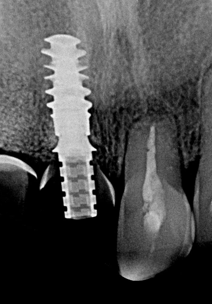
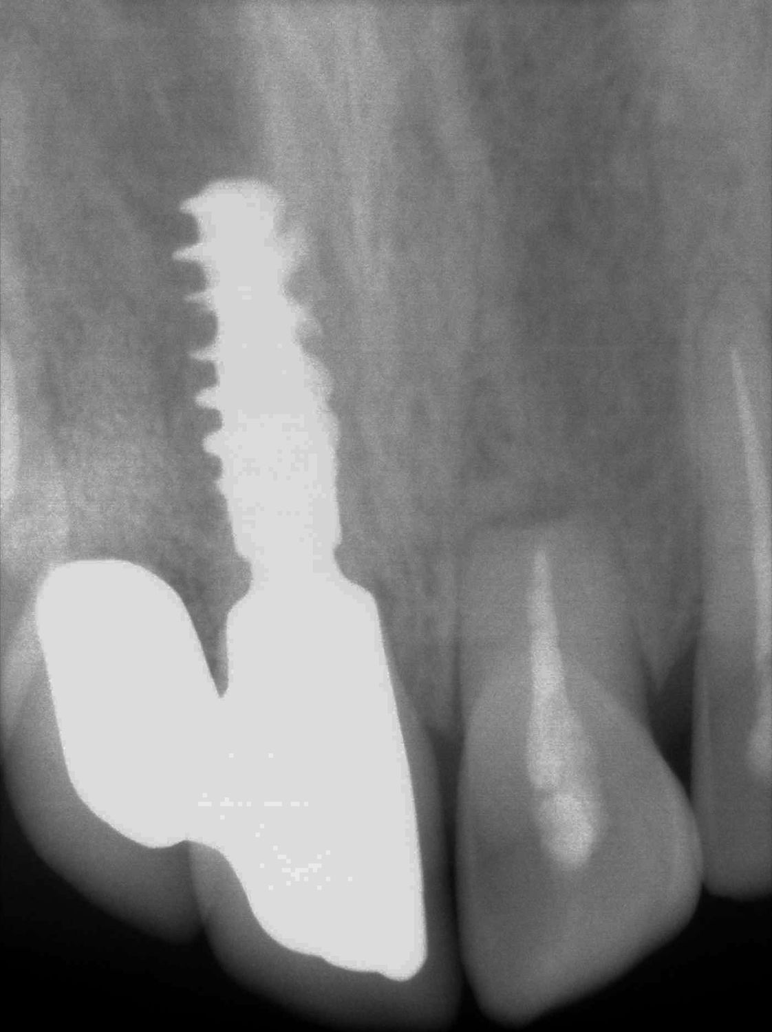
The clinical and radiological exams showed no signs of bone or adjacent teeth fracture. Nevertheless, we decided to take a CBCT for better planning of the case. In fact, bone plates were intact. Still, buccal bone plate showed thin and fragile. In cases like this, apical anchorage is the only way to increase primary stability. (Figs. 2-3) First approach was a removable partial denture made up with ovate pontics, as to preserve soft tissue shape. To increase primary stability, Straumann® BLX implant was the best option. With its unique functional design, it provides simplicity and immediacy. Progressive thread design compacts bone, increases its density and thus, increases strength. It cuts, collects, and condenses native bone, achieving almost perfect BIC (bone-to-implant contact).

For better management of soft tissues, we decide a Type 2A, early placement and immediate restoration loading, 4 weeks after the incident.

Surgical procedure

First, a full thickness mucoperiosteal flap was elevated, and confirmed the presence of buccal plates. (Fig. 4)
Re-directing of drilling and implant placement ensures avoiding socket apex and entering the palatal plate. This leaves a gap between implant and buccal plate which can be filled with slow resorption bone graft. Straumann® VeloDrill™ System, and patient’s denture was used as a surgical guide. (Figs. 5-6) Parallelism pin, and radiographic control. (Figs. 7-8)
A Straumann® BLX 4.5 ø x 10 mm implant was placed, slightly palatal inclination, into the upper right central incisor’s socket, with an optimal insertion torque. Implant per se doesn’t secure optimal esthetic results. It is mandatory to use biomaterials if we mean to maintain bone contour and gain gingival width and thickness.
The gap between implant and buccal plate was filled with Cerabone® —which is bovine bone substitute — and, seeking for a contour augmentation, a Mucoderm® acellular collagen matrix was placed and fixed with stitches. (Figs 9-10)
Flap adaptation, and tension free primary closure. (Fig. 11)
Same removable provisional denture was adapted and adjusted to be modified as a fixed restoration and used for an interim prothesis. (Fig. 12-13)


Want to stay up to date?

youTooth.com is THE PLACE TO BE IN DENTISTRY – subscribe now and receive our monthly newsletter on top hot topics from the world of modern dentistry.


Prosthetic procedure

Three months later, with the presence of a mature and healthy peri-implant tissue, provisional restoration was modified twice, in order to improve emergence profile and the conformation of our ovate pontic tissue. We used a chemical-restorative combination of corticosteroid and lytic enzymes, to achieve faster re-epithelization and repeated after 2-3 weeks until tissue was ready.

On subcritical contour, a concave shape was molded and improved with fluid resin, to gain as much soft tissue as possible. (Figs. 14-15)

Then a radiograph was taken to visualize temporary abutment and fluid resin modifications made on contour. (Fig. 16)

Final contour was established (Fig. 17), having 5 mm from contact point to abutment base.

Consistent shape of all Straumann® abutments, from healing cap, temporary coping, to SRA or Variobase® provides a definitive and constant diameter for junctional epithelium fibers and approaches the one abutment one time philosophy.

Five months after surgery, almost 5 mm were accomplished from abutment base to papilla tip. (Fig. 18) Impression coping was personalized (Fig. 19-21) and an open tray impression technique at implant level was performed (Fig. 22-23).

After impression, color matching was made. (Fig. 24-25)

For the definitive prothesis, mono scan body was acquired by digital workflow on a dental laboratory. Milled monolithic splint ZrO2 crowns with cantilever, characterized with cut back technique, on a Variobase® 3.5mm abutment height, 1.0mm gingival height. Hybrid cemented-retained and screw-retained technique, sandblasting and cemented with anaerobic resin-based cement (Figs. 26-29)

Definitive crowns were tried on cast, which had an almost perfectly accurate reproduction of gingival margins. (Fig. 30)

Front and 45 angle pictures showed an interproximal papillae with a very natural-looking contour and an excellent esthetic result. (Figs. 31-32)

An immediate radiograph was taken to control definitive prothesis adjustment. (Fig. 33)

Treatment outcomes

Six months clinical and radiographic follow up, shows desirable esthetic results regarding gingival contour and interproximal papillae. (Fig. 34) One of the keys in modern implant rehabilitation is to shorten treatment time. In five months, patient had its definitive crowns, and his chief complaint was addressed successfully.

Straumann® BLX has been developed to improve long term predictability and the use of immediateness as an option in daily implementation. Also providing stability, versatility, confidence, and innovation. It allows us to develop and capitalize new business opportunities, approaching the increasing demand on short term treatments.

References:

  1.  Spear Frank. The use of implants and ovate pontics in the esthetic zone. Compendium March 2008, Vol.29 Number 2.
  2. Tarnow D, et al. Vertical distance from the crest of bone to the height of the interproximal papilla between adjacent implants. J Periodontology 2003 Dec;74(12): 1785-1788. 
  3. Lundgren D, et al. Biomechanical aspects of fixed bridgework supported by natural teeth and endosseous implants Periodontology 2000 Vol.4, 1994:23-40. 
  4. Wittneben JG, Millen C, Bräger U. Clinical performance of screw versus cement-retained fixed implant-supported reconstructions. A systematic review. 5to ITI Consensus Conference. Int J Oral Maxillofac Implants 2014;29(Suppl):84-98.
  5. Sailer Irena, et al. Cemented and screw-retained implant reconstructions: a systematic review of the survival and complications rates. Clin Oral Implants Res 23 (suppl 6), 2012, 163-201.