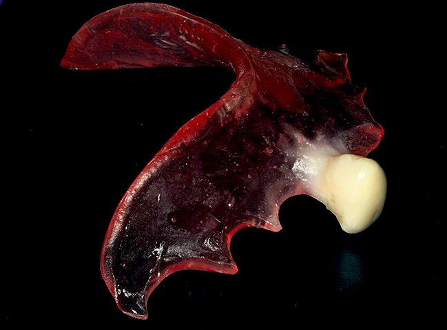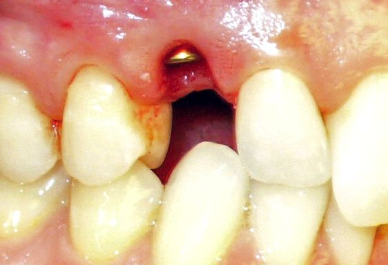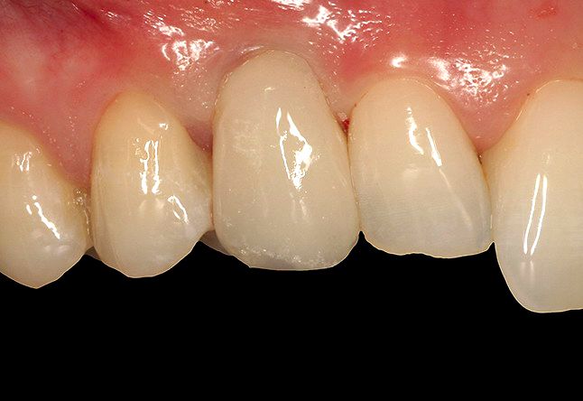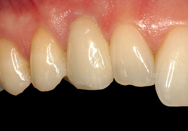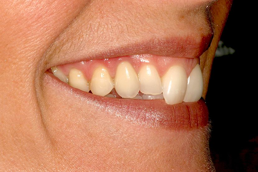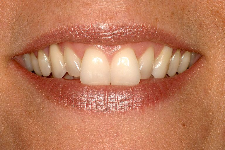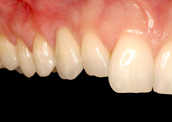

A 39-year old female with non-remarkable medical history presented for extraction of the non-restorable primary maxil- lary right canine (53) and implant replacement (Figs. 1–3). Tooth in Regio 13 had been removed over 20 years ago because of high impaction.
Treatment planning
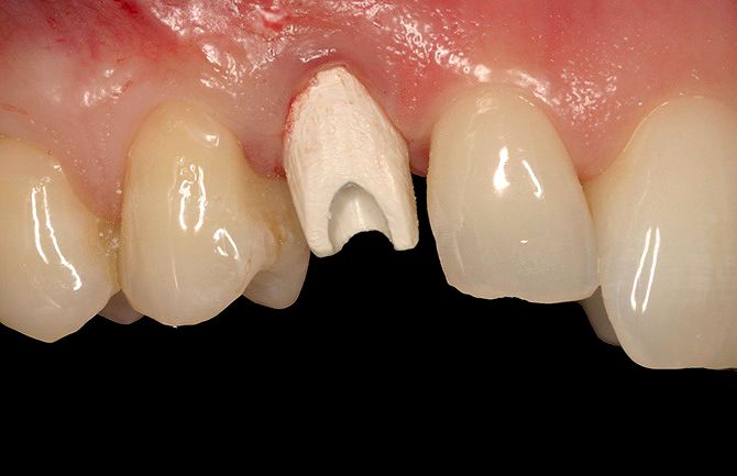
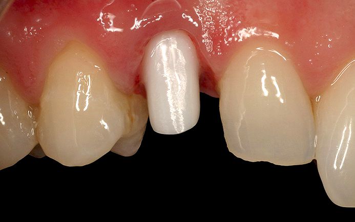
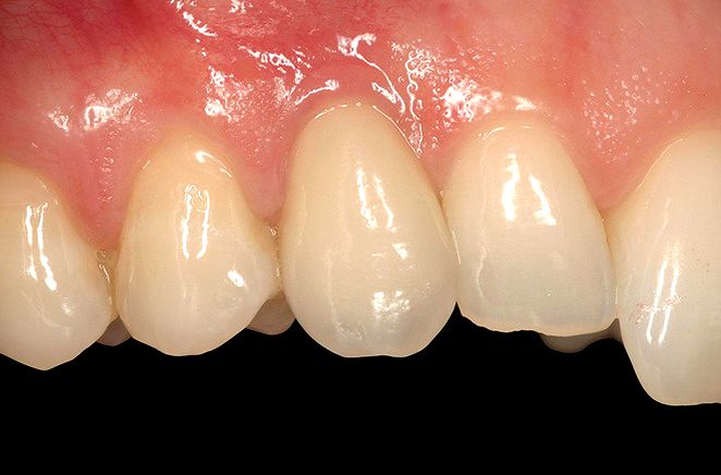
A delayed implant placement was planned to achieve a better control of the soft tissue contour and architecture. The alveolar ridge was narrow with buccal undercut, therefore, a small diameter implant design was treatment planned for the site. The potential for high forces, compromised bone quality and quantity, as well as esthetic demands required the use of a fast healing and strong implant with prosthetic flexibility to achieve the desired goal.
Surgical procedure
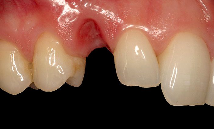
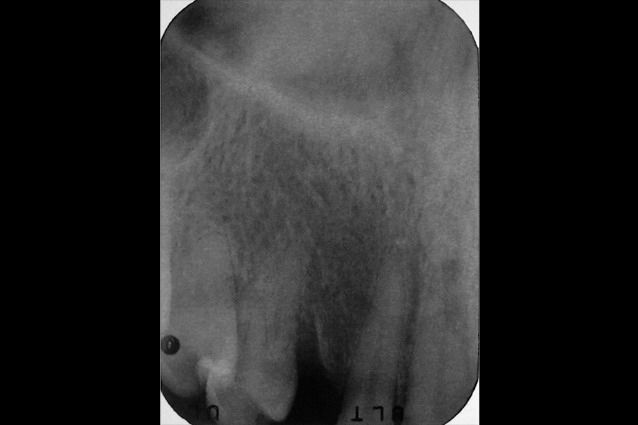
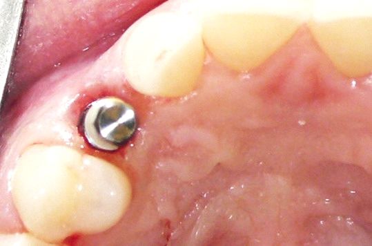
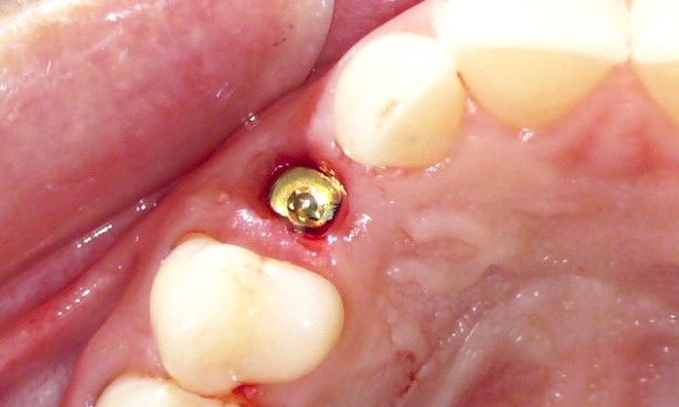
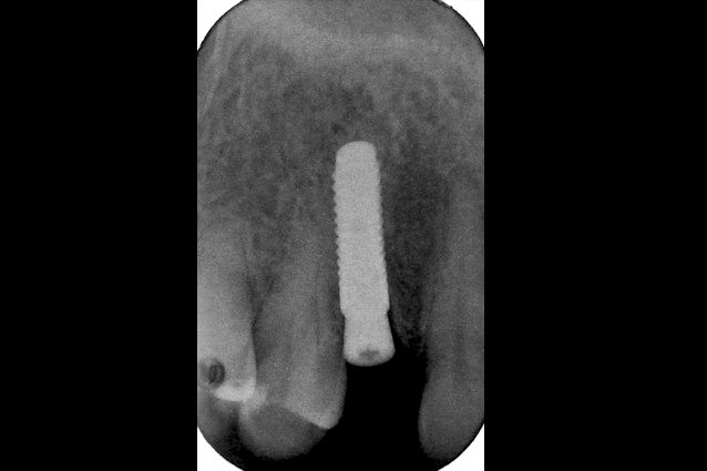
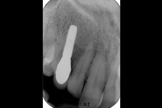
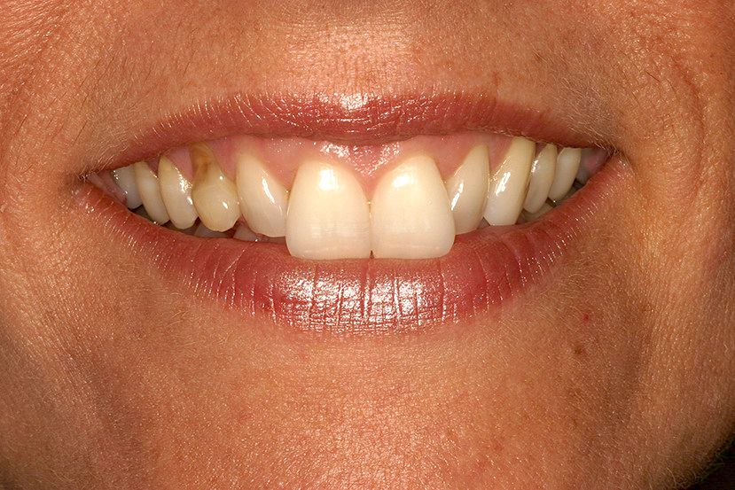
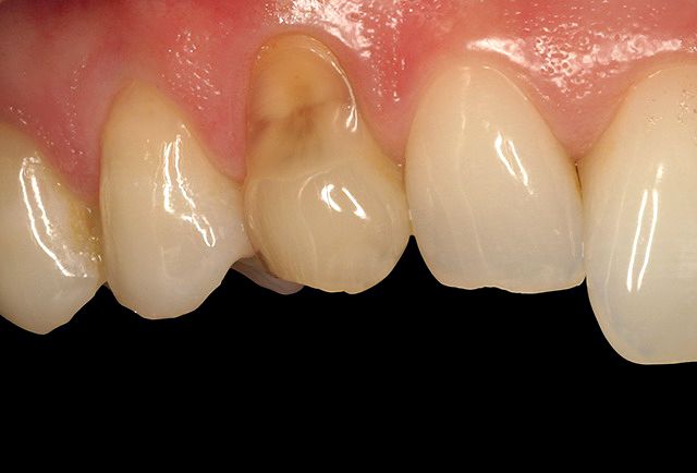
The initial phase involved removal of tooth in Regio 53 and placement of an interim removable partial denture to provisionalize the site (Fig. 4). The denture tooth in the prosthesis was shaped as an ovate pontic to contour the gingiva (Fig. 5) while awaiting implant placement (Fig. 6). Two weeks later, a flapless osteotomy with cooled saline irrigation was made in type 3 bone, following the socket contours to help pre- serve the original canine eminence. During the osteotomy, a buccal fenestration was created apical to the socket of the primary tooth. After completion of the osteotomy using a final drill of 2.8 mm, the fenestration was grafted with a xenograft material through the osteotomy. A Roxolid® implant (Straumann® Bone Level Implant NC 3.3 mm, SLActive® 12 mm) was placed with primary stability using a hand- piece at 35 Ncm (Figs. 7, 8). A conical healing abutment (Ø 3.6 mm/height 3.5 mm) was placed and no sutures were required (Figs. 9–11).