
Introduction
A determinant of long-term implant success also lies in the selection of an appropriate implant system. Straumann® BLT implants, characterized by their proprietary Roxolid® material and SLActive® surface, have garnered considerable attention for their osseointegration potential and stability1,2. These implants mimic a dental root shape, as they have a smaller diameter at the apical part than at the neck of the implant. The claimed benefits of this design include enhancement of the primary stability by the pressure of the cortical bone on regions with poor bone quality, as well as the reduced risk of bone perforation due to its macrotopography3.
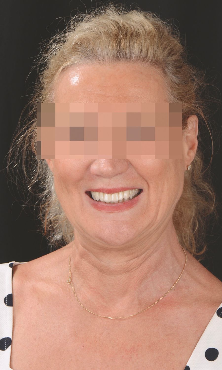
This case report presents the 9-year follow-up of two Straumann® BLT implants placed in the esthetic zone, focusing on their clinical performance, peri-implant tissue health, and patient satisfaction. By examining the longevity and esthetic outcomes, this report highlights the importance of careful treatment planning and execution in achieving predictable outcomes. Initial situation A 56-year-old female patient, non-smoker, classified as healthy (ASA I), with no current medications or known allergies, visited our clinic with a chief complaint centered around her persistent dissatisfaction with her smile. She reported the development of a chronic infection in her front teeth over recent years, leading to noticeable mobility. This dental concern has significantly affected her ability to eat and speak with confidence. The patient was actively seeking a long-term solution but expressed concerns about potential pain during the treatment process. The patient's extraoral examination revealed a medium smile line and misalignment of the front teeth (Figs. 1,2).
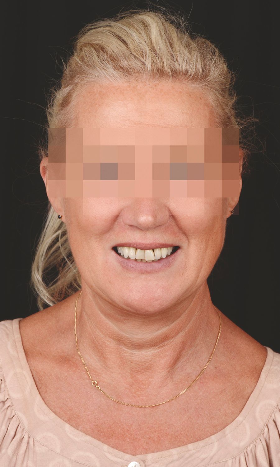

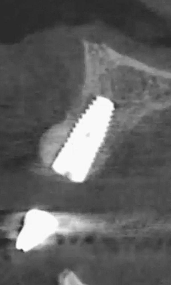
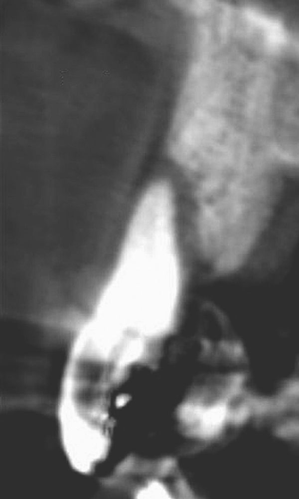
During the intraoral examination, advanced periodontal attachment loss and mobility were noted in teeth #12, #21, and #22 (Fig. 3). Cone-beam computed tomography (CBCT) imaging indicated the absence of buccal bone on tooth #21 (Fig. 4).

According to the SAC classification, the patient was classified surgically as complex and prosthetically as straightforward (Fig. 5).