#Esthetics 30. Mar 2023

Interdisciplinary approach for a missing upper incisor: Orthodontic treatment, GBR & placement of a Straumann® BLX implant

A clinical case report by Tran Hung Lam, Vietnam

Implant therapy aims to provide patients with a highly predictable treatment outcome, good long-term stability, and a low risk of complications during the healing and follow-up phases. The growing demand to restore missing teeth in the functional and esthetic aspects has become an important challenge. This is especially true in the anterior zone, as various local risk factors can compromise the predictability of the results. Therefore, the clinician must carefully examine the patient's risk profile before establishing the treatment plan.(1)

Introduction

The ITI recommends immediate implant placement (type 1) in the presence of ideal anatomic conditions. This includes (i) a fully intact facial bone wall with a thick-wall phenotype (> 1 mm) at the extraction site, (ii) a thick gingival biotype, (iii) no acute infection at the extraction site, and (iv) a sufficient volume of bone apical and palatal to the socket to allow implant insertion in a correct 3D position with sufficient primary stability. When these ideal conditions are not met, it is suggested to place implants after 4-8 weeks of soft tissue healing (type 2). If primary stability cannot be achieved after 4-8 weeks, the post-extraction healing period should be extended to allow for partial bone healing (type 3).1 Type 4 is the placement of the implant into a fully healed site.2

An adequate amount of bone is needed to be able to place the implant in an ideal prosthetic-driven position. If there adequate bone volume is not available, guided bone regeneration (GBR) techniques should be used for ridge augmentation before implant placement.3

The following case report describes an interdisciplinary treatment that included orthodontic therapy, guided bone regeneration (GBR), implant placement, and fixed restorations. Ceramic braces were used to level, align, and gain space for implant placement at a central incisor location. Because of the complexity of this clinical case, the GBR was first carried out with a non-resorbable membrane and Cerabone® and, after six months, a Straumann® BLX implant was placed.

Initial situation

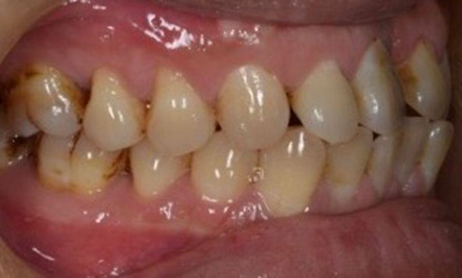
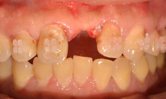
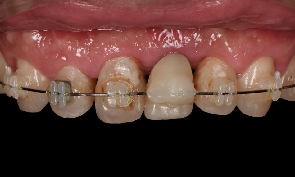
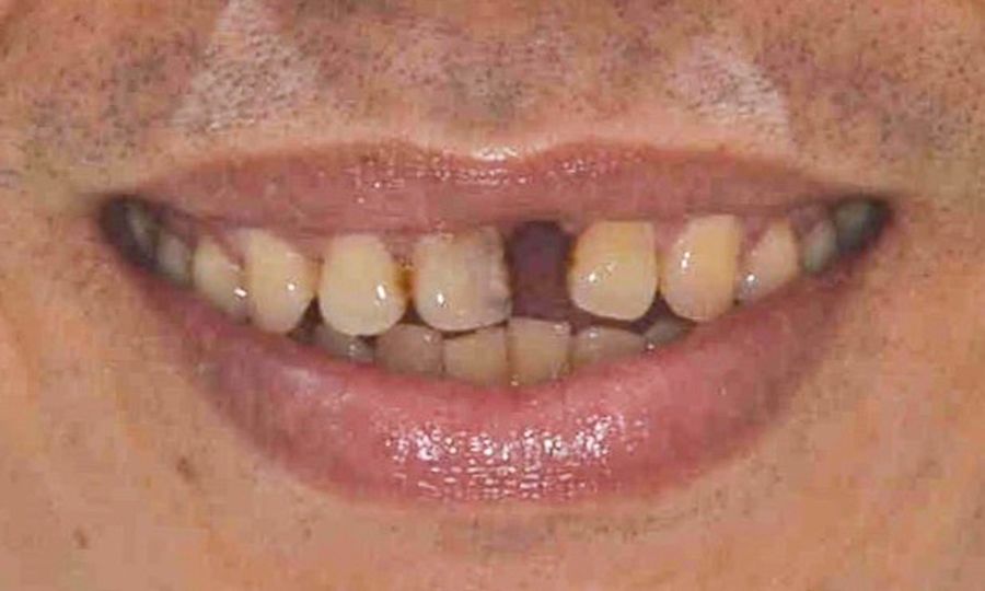
A systemically healthy 48-year-old male patient came to our clinic seeking an esthetic and functional treatment for a missing anterior tooth. He reported being a non-smoker, taking no medication, and with no allergies. His chief complaint was, “I feel very embarrassed to talk and smile in public because I have a missing tooth. I would like to have a fixed restoration and a nice smile. I don’t want to wear this denture any longer.” His dental history revealed the loss of tooth #21 during an accident over 20 years ago. Since then, he had noticed that the space left by the central incisor was slowly being closed by the adjacent teeth. The extraoral examination revealed a medium smile line with an impaired mesiodistal (MD) proportion of the anterior teeth. Due to the limited MD availability on position #21, the provisional restoration looked small and narrow. Moreover, the anterior teeth were not level, resulting in a reverse smile. For the intraoral examination, the provisional denture was removed. Neighboring teeth were mesially tilted (Fig. 1).

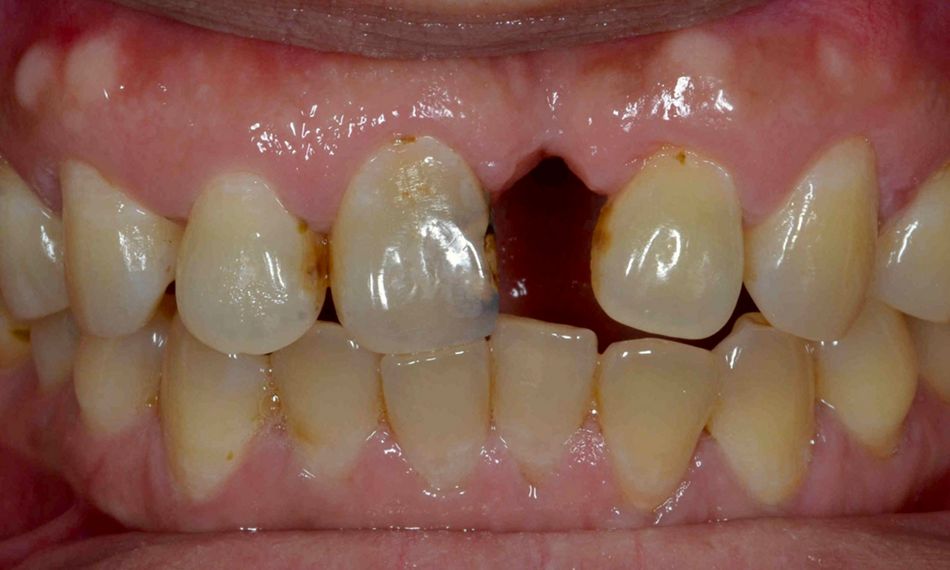
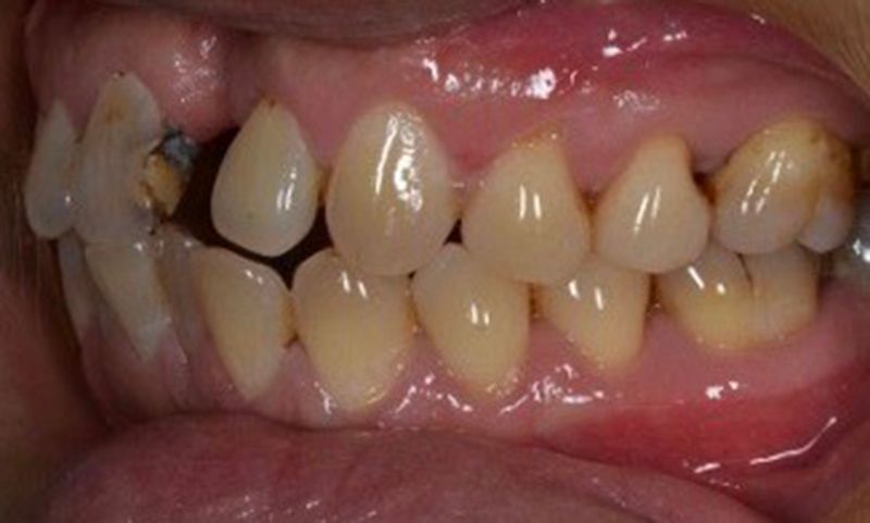
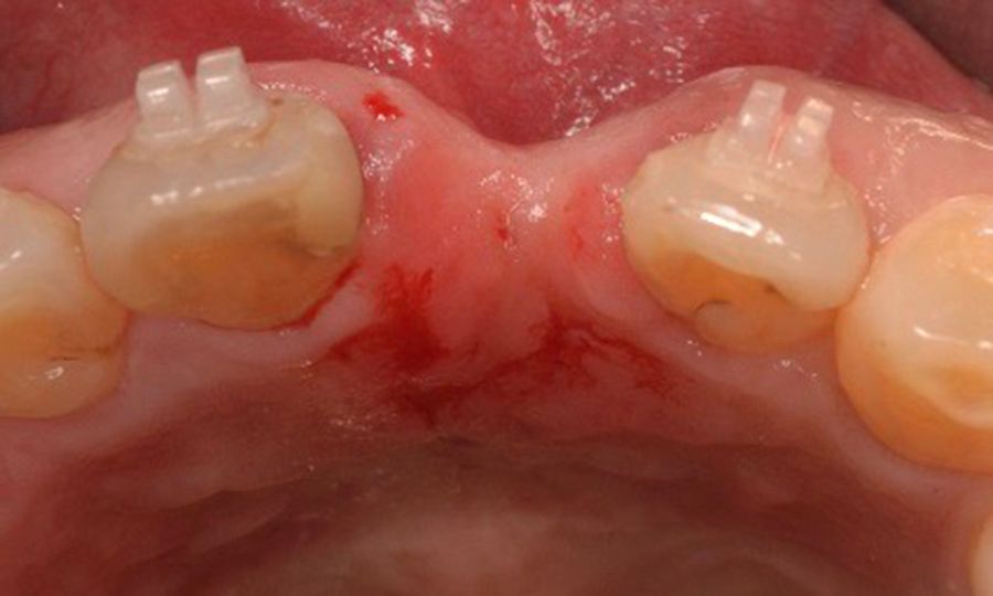
Since the residual ridge was atrophic, a severe horizontal ridge defect was apparent, and secondary caries was present in #11 distally (Figs. 2-4).

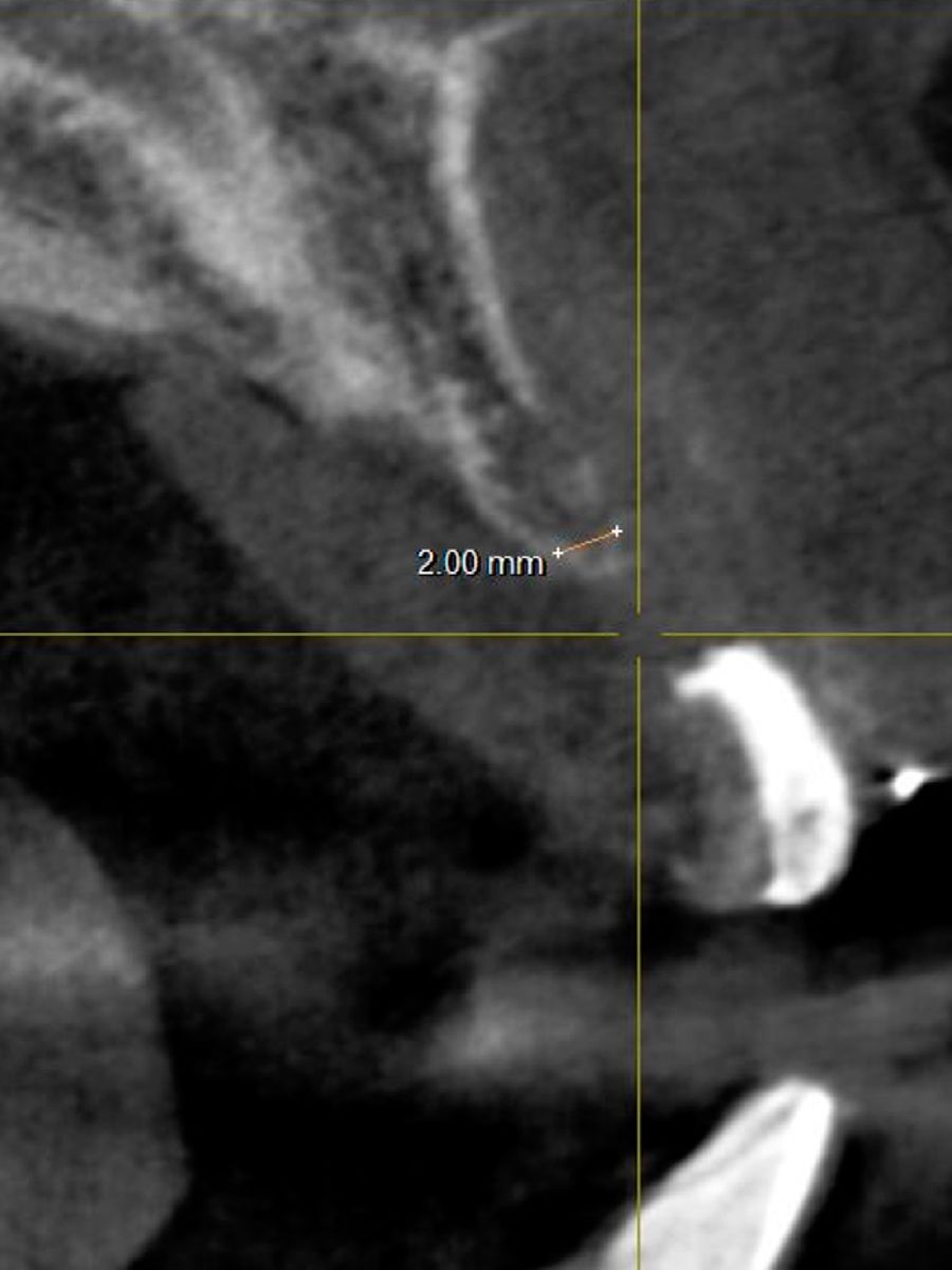
The radiographic assessment (CBCT) revealed a narrow crestal width at site #21 and no local infection (Fig. 5).

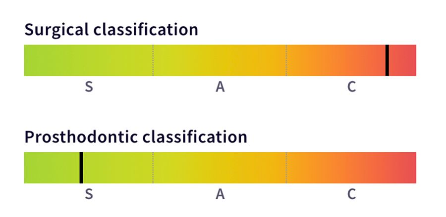
The SAC classification assessed the potential difficulty, complexity, and risk of the implant-related treatment. The patient was classified as complex surgically and straightforward for prosthodontics (Fig. 6).

After evaluating the patient's wishes and discussing the treatment options, it was decided first to perform orthodontic treatment, then GBR and, finally, the placement of a Straumann® BLX implant. The Straumann® BLX Implants are made from the material Roxolid® with the SLActive® surface. These unique properties enable enhanced control over insertion torque to achieve optimal primary stability, which was a fundamental feature in treating this type of clinical scenario.

Treatment planning

1. Oral hygiene instructions and non-surgical periodontal treatment.
2. Digital planning of dental space distribution and esthetics.
3. Caries restorations and orthodontic treatment to increase the mesiodistal gap at site #21 and to level and align the smile curve (Figs. 7,8).

4. Guided bone regeneration using a non-resorbable membrane and Cerabone®
5. Membrane removal after six months and Straumann® BLX implant insertion in a prosthetic-driven position.
6. Temporary screw-retained crown delivery.
7. Crown preparation on tooth #11.
8. Definitive screw-retained crown delivery.

Surgical procedure

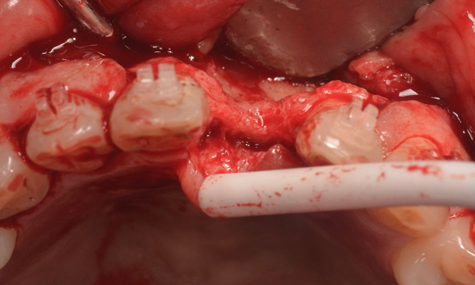
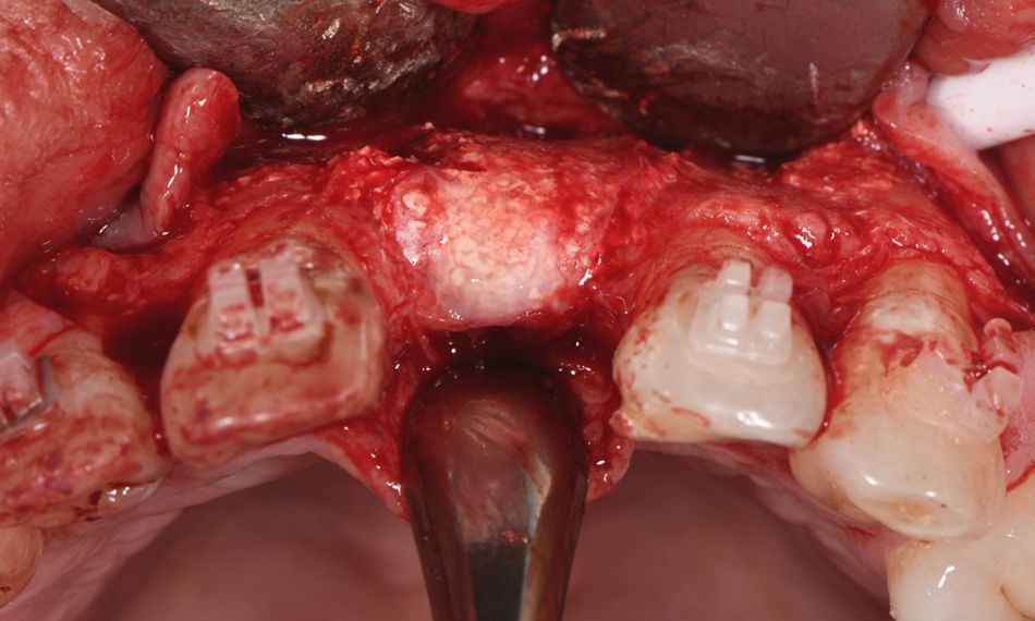
Due to the limited bone availability, the first step of the surgical procedure was the guided bone regeneration using a non-resorbable membrane and Cerabone®. Lidocaine 2% with epinephrine 1:100k was administered, and a mucoperiosteal flap with a crestal incision was raised. The flap was carefully separated from the bone, and the surgical access confirmed the limited availability of bone (Fig. 9).

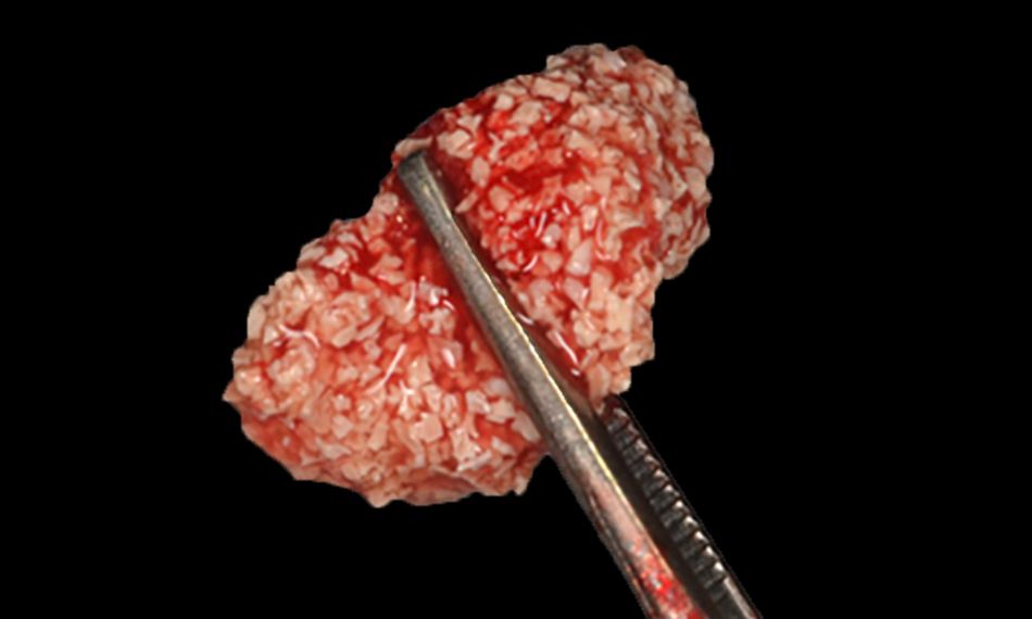
Afterward, guided bone regeneration was performed. Cerabone® was used as a bone substitute, which is bovine bone grafting material. In addition, a non-resorbable membrane to exclude non‐osteogenic tissues from interfering with bone regeneration was used (Fig. 10).

The patient was advised to have a soft diet and use ice packs in the area during the first 48 hours. Moreover, the postoperative prescription included rinsing with an antiseptic solution (for 1 minute with chlorhexidine 0.2% twice a day for one week), painkillers (ibuprofen 600 mg up to four times a day as required), and antibiotics (amoxicillin 500mg three times a day for 5 days).

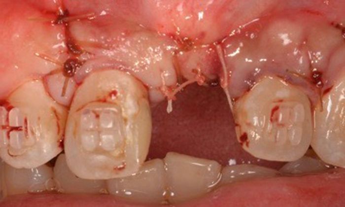
Two weeks later, at the suture removal appointment, the healing was uneventful, and the patient reported no complications.

The patient returned six months after surgery for a follow-up evaluation. The healing and oral hygiene were good. Furthermore, there was an adequate mesiodistal gap at position #21 for implant placement, thanks to the orthodontic treatment (Fig. 11).

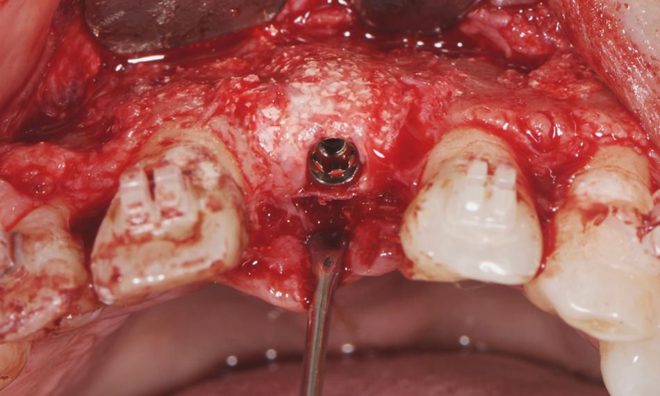
The implant placement was planned. Following local anesthesia infiltration, the area was re-opened with a full-thickness flap for membrane removal. The bone morphology and dimensions were assessed and found to be optimal for implant insertion (Fig. 12).

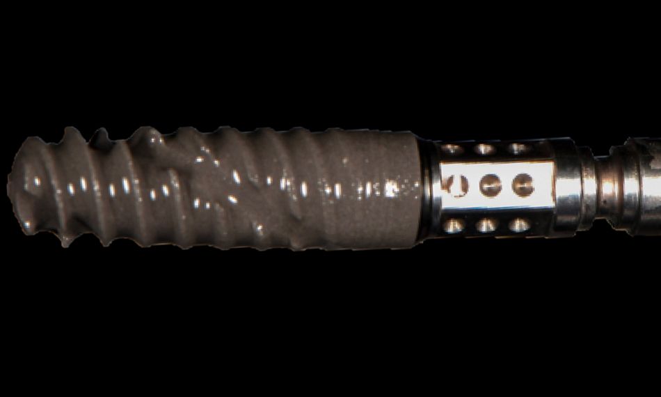
The Straumann® BLX Implant 3.75 mm SLActive® 12 mm Roxolid® was selected (Fig. 13). The surgical bed was prepared, and the implant was placed in a prosthetic-driven position following the manufacturer's instructions (Fig. 14). Next, the mucoperiosteal flap was adapted and closed with interrupted sutures, achieving primary closure (Fig. 15).

At the suture removal appointment, since healing was uneventful, the braces were removed, and a screw-retained temporary restoration was delivered (Figs. 16,17).

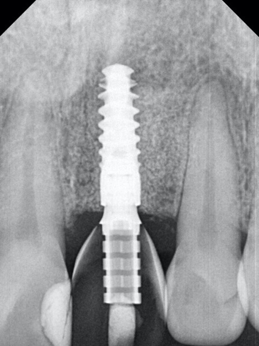
A periapical radiograph was taken to assess the correct fit of the restoration (Fig. 18).

Prosthetic procedure

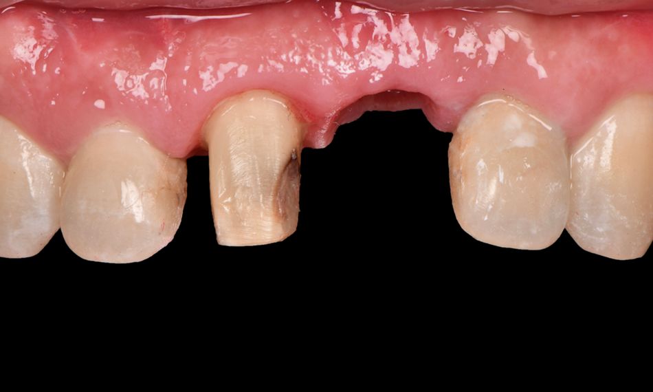
Twenty weeks after implant surgery, the papillae were well conformed. In addition, crown preparation was performed on tooth #11 (Fig. 19).

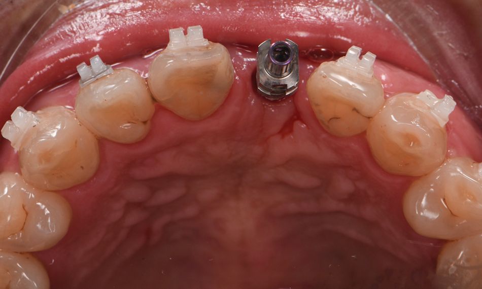
Osseointegration was achieved on #21, and the Straumann® RB Variobase® and zirconia coping obtained by the CAD/CAM procedure for the final restoration of the BLX implant were placed (Fig. 20).

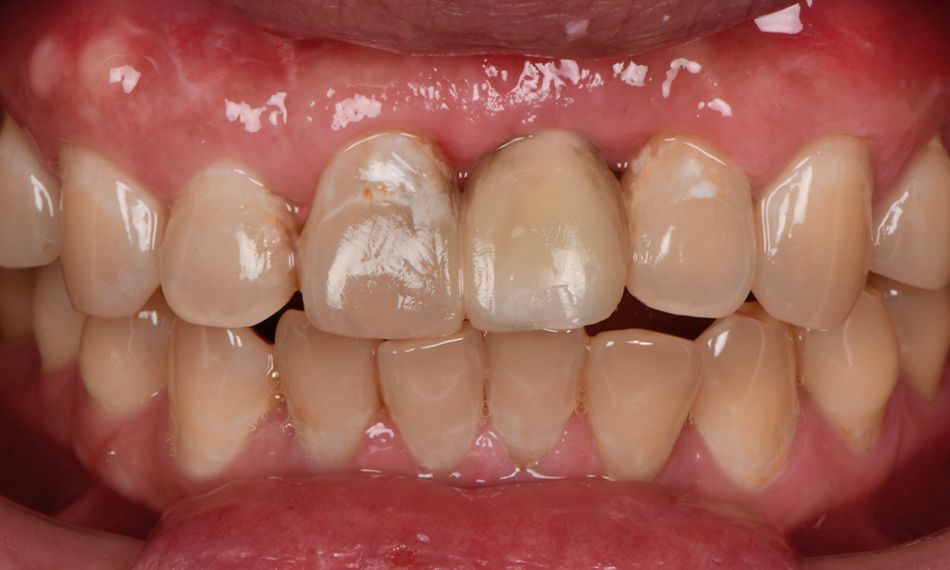
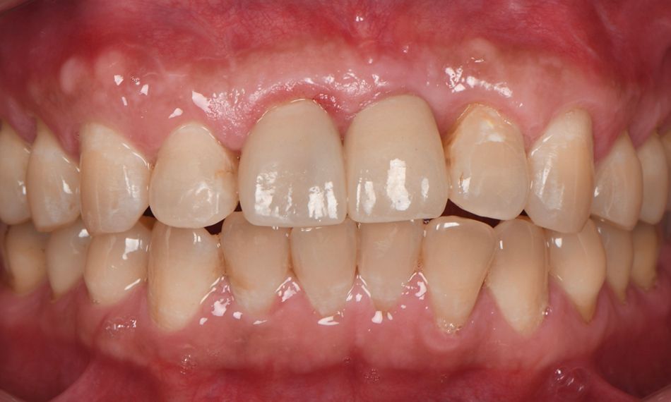
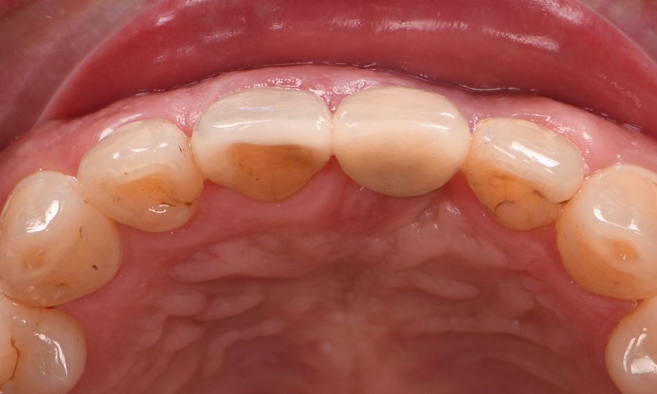
The final implant restoration was done at position #21, and a lithium disilicate crown was placed on tooth #11 (Fig. 21). The occlusal view shows a natural contour of soft and hard tissues (Fig. 22). Occlusion was checked, and oral hygiene instructions were reinforced.

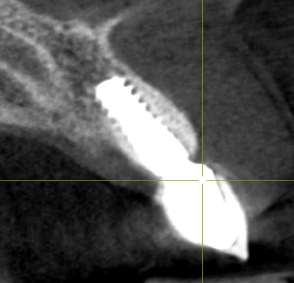
The patient was involved in an annual maintenance program in which soft and hard tissues are evaluated and oral hygiene instructions reinforced. The radiographic control after three years shows good maintenance of the peri-implant bone (Fig. 23).

Treatment outcomes

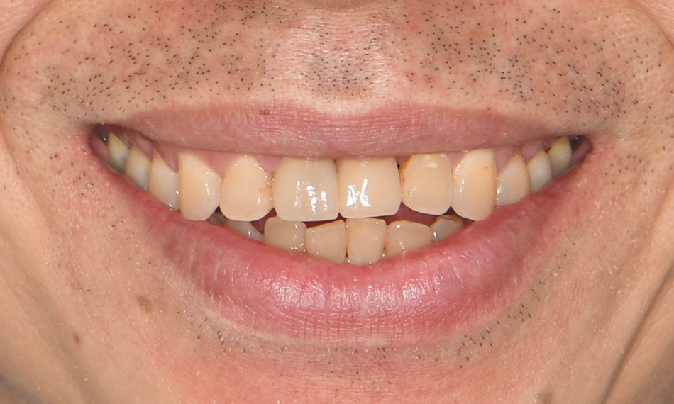
The outcome met our patient’s expectations. In addition, the hard and soft tissues were well maintained over time (Fig. 24).

Recently, during the 3-year follow-up visit, the patient stated: “This treatment has greatly impacted my life; I regained my confidence and self-esteem. I love my new smile, and everyone notices it because I've never smiled so much.”

Author’s testimonial

In my daily practice, the use of Straumann® BLX implant on sites that previously underwent guided bone regeneration is key for the success of the implant treatment. The BLX implant design allows optimal primary stability without pressure on the regenerated crestal bone.

References:

  1. Buser, D., Chappuis, V., Belser, U.C. and Chen, S. (2017), Implant placement post extraction in esthetic single tooth sites: when immediate, when early, when late?. Periodontol 2000, 73: 84-102.
  2.  Morton D, Chen ST, Martin WC, Levine RA, Buser D. Consensus statements and recommended clinical procedures regarding optimizing esthetic outcomes in implant dentistry. Int J Oral Maxillofac Implants. 2014;29 Suppl:216-20. doi: 10.11607/jomi.2013.g3. PMID: 24660199.
  3. Eswaran B, Rai R, Rathee S, Chirom B, V M. Aesthetic essentiality regained using implant- a case report. J Clin Diagn Res. 2014;8(8):ZD01-ZD3.