#Full-Arch 3 d ago

Full-arch rehabilitation using Straumann® Roxolid® ⌀3.3 mm implants with Straumann® RevEX™ workflow: Optimizing precision, esthetics, and function

A clinical case report by Dr. José Manuel Abarca, Dr. María José Bernedo, Dr. Pablo Cavalerie and Dr. Javiera Aguilera - Chile

Full Arch Rehabilitation using Straumann®️∅3.3 mm implants, GBR and RevEX™️ Workflow

Introduction

Digital dentistry has gained popularity and become a routine part of clinical practice, encompassing areas such as digital planning, guided surgery, and rehabilitation with fixed prostheses and implants. Intraoral scanners, which offer a three-dimensional preview of prostheses with equal or better accuracy than conventional techniques, have improved the patient experience and optimized clinical time for dentists.

However, the digitization of completely edentulous ridges for implant-supported full-arch rehabilitation remains challenging, especially in the mandible, due to several technical and anatomical issues. To address these challenges, a solution known as the reverse scanbody has been developed. This innovative concept allows for accurate records of implant locations using extraoral scans of provisional prostheses.

This case report highlights a full-mouth rehabilitation utilizing six Straumann® BLT ∅3.3 mm SLActive® Roxolid® implants and a digital workflow with Straumann® RevEX™ scan body. This comprehensive approach effectively addressed the patient’s functional requirements while exceeding esthetic expectations, resulting in a significant enhancement of confidence and overall quality of life.

Initial situation

A systemically healthy 42-year-old male non-smoker consulted for a definitive prosthetic solution.

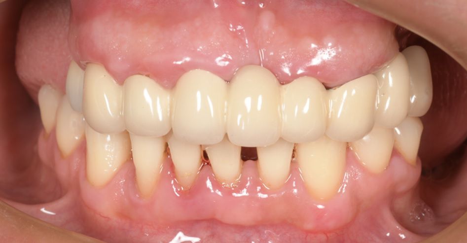
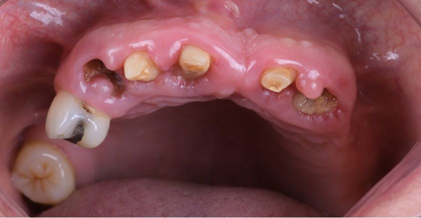
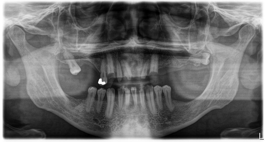
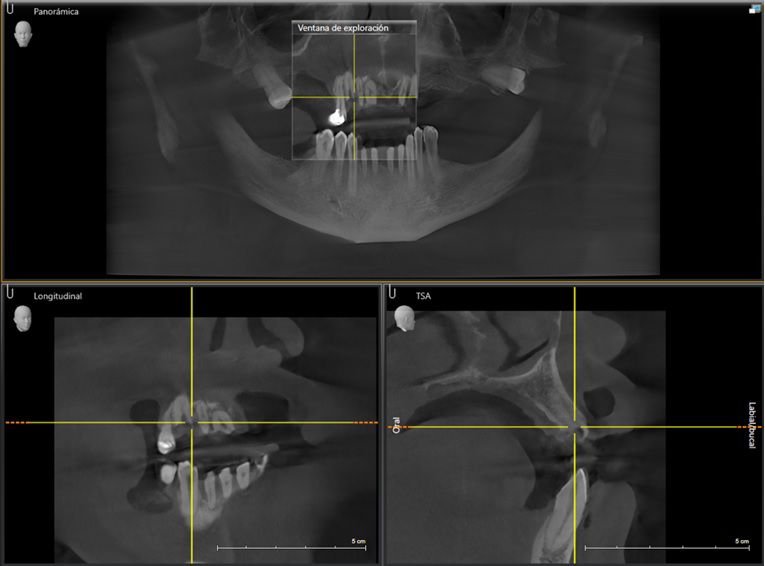
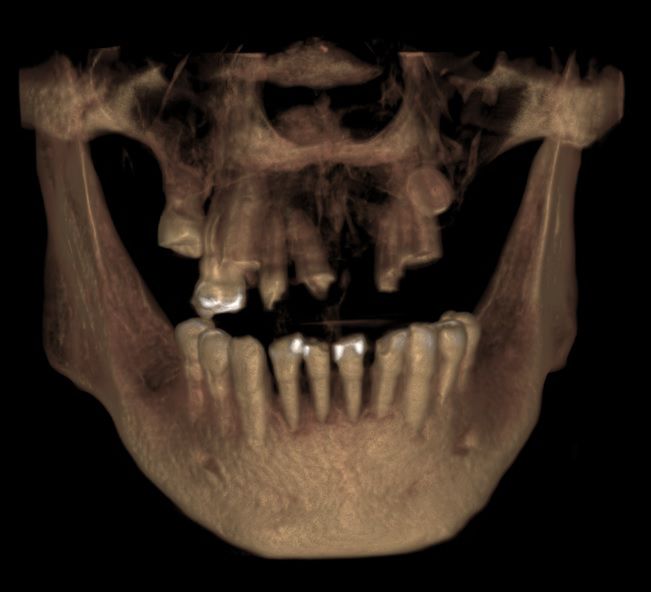
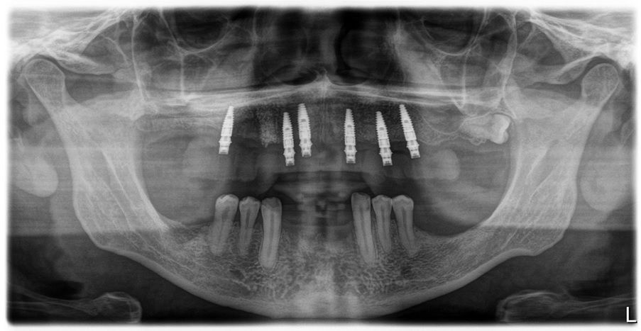
An intraoral clinical examination showed an upper plural fixed prosthesis spanning from #13 to #23, with coronal remnants lost due to secondary caries, an unfavorable prosthetic prognosis, and gingival inflammation associated with prosthetic misalignment. Additionally, there was significant horizontal and vertical bone resorption in sextants 1 and 3 (Figs. 1-5).

Treatment planning and surgical procedure

A different therapeutic approach was planned for the maxilla and mandible. For the maxilla, a full-arch implant-supported prosthesis was planned, while a removable partial denture was chosen for the mandible.

An initial cause-related therapy was performed, including scaling and multiple extractions of the remaining upper teeth. The procedure was conducted under local anesthesia using 3 × 1.8 mL cartridges of 4% articaine with 1:100,000 epinephrine. Nonsteroidal anti-inflammatory drugs were prescribed for pain management, and a follow-up appointment for control and suture removal was scheduled 12 days post-surgery.

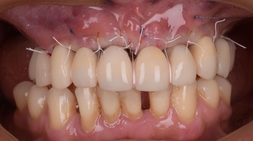
During maxillary surgery, vestibular and palatal anesthesia infiltrations were administered, and crestal and intrasulcular incisions were made with a 15C scalpel to create a flap, allowing for dental extractions and minimally invasive exposure of the surgical field. Teeth #17, #15, #13, #12, #11, #22, and #23 were carefully removed to minimize bone loss.

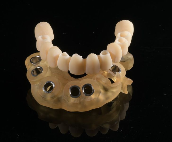
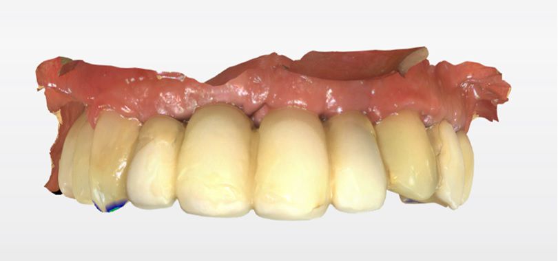
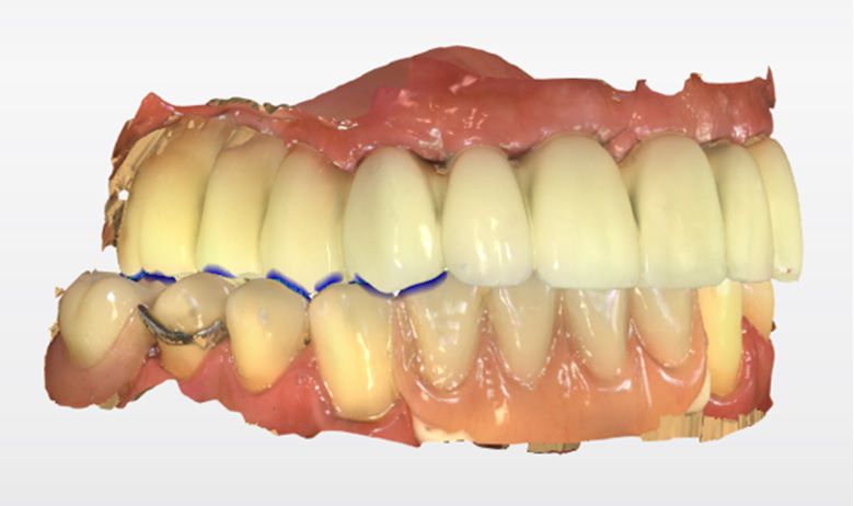
Digital records were obtained for prosthetic planning, including the creation of a surgical guide and the fabrication of a 3D-printed temporary upper prosthesis (Figs. 6,7). A lower temporary prosthesis was also planned to replace the teeth #31, #32, #41, and #42.

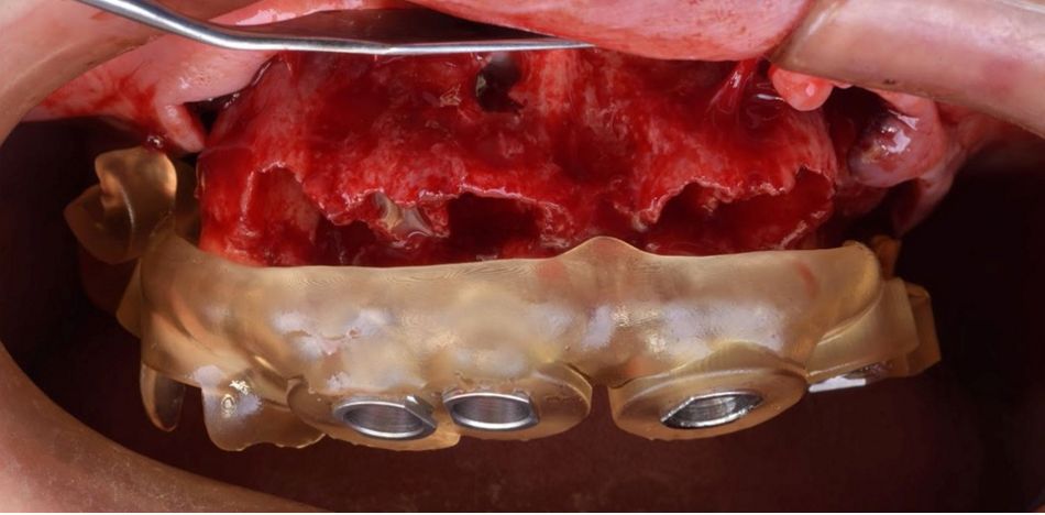
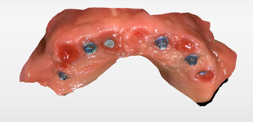
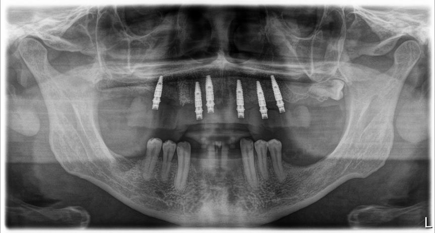
Guided placement of six Straumann® BLT ∅3.3 mm NC, SLActive® 12 mm Roxolid® implants was performed using the Straumann guided surgery protocol. Each implant achieved adequate initial stability, exceeding 35 Ncm of torque. Subsequently, Straumann® screw-retained Ø3.5 mm abutments were secured on each implant at a torque of 35 Ncm, as recommended. Additionally, supplemental augmentation surgery was performed at sites with deficient bone using guided bone regeneration with cerabone® (bone substitute) and a Jason® membrane, which was secured with a subperiosteal suture. The printed temporary prosthesis was then prepared by cementing six Variobase® for SRA abutments (Figs. 8,9).

Once in place, a palatal-harvested connective tissue graft was secured to the buccal flap, and a sling suture was used to promote proper esthetic healing.

After eight weeks, definitive loading was performed with a cement-retained zirconia prosthesis placed on Variobase® abutments for SRA. This was achieved using a 100% digital workflow with the Straumann® RevEX™ scanbody system as the scanning tool.

Prosthetic procedure

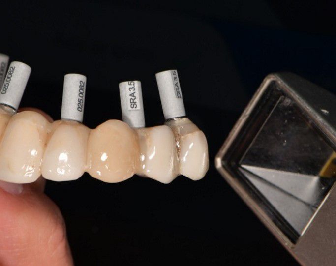
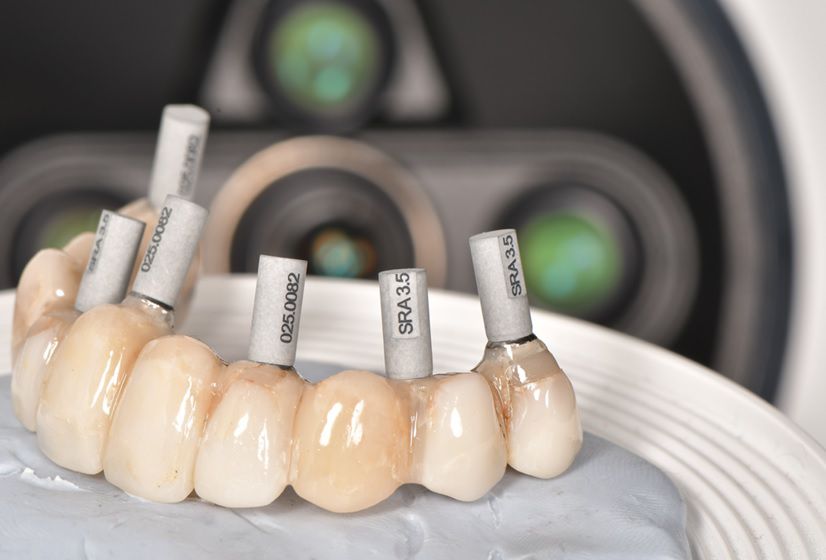
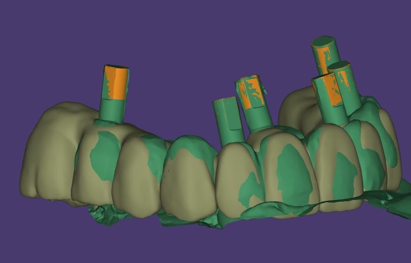
Once the osseointegration period was complete, the final digital impression was taken with the Straumann® RevEX™ reverse scanbodies placed in the final temporary restoration. Three scanning methodologies were used to compare the accuracy and clinical performance of the resulting rehabilitations (Figs. 10,11).

1. Intraoral scanner and provisional held manually.
2. Intraoral scanner of provisional with helix pattern on a mobile platform.
3. Desktop scanner.

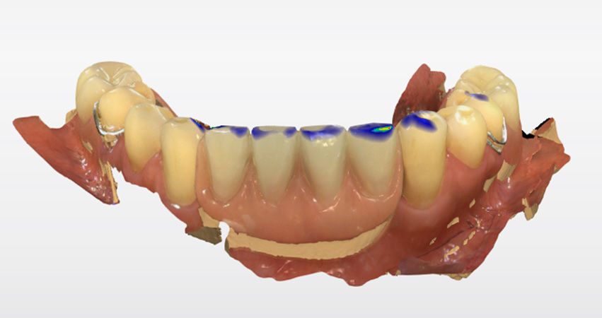
The protocol also included digitally recording the antagonist arch and the maxilla with the screw-retained provisional prosthesis in place. Next, the occlusal registration was scanned, followed by a scan of the maxillary soft tissue with the six Straumann® screw-retained abutments (Figs. 12-15).

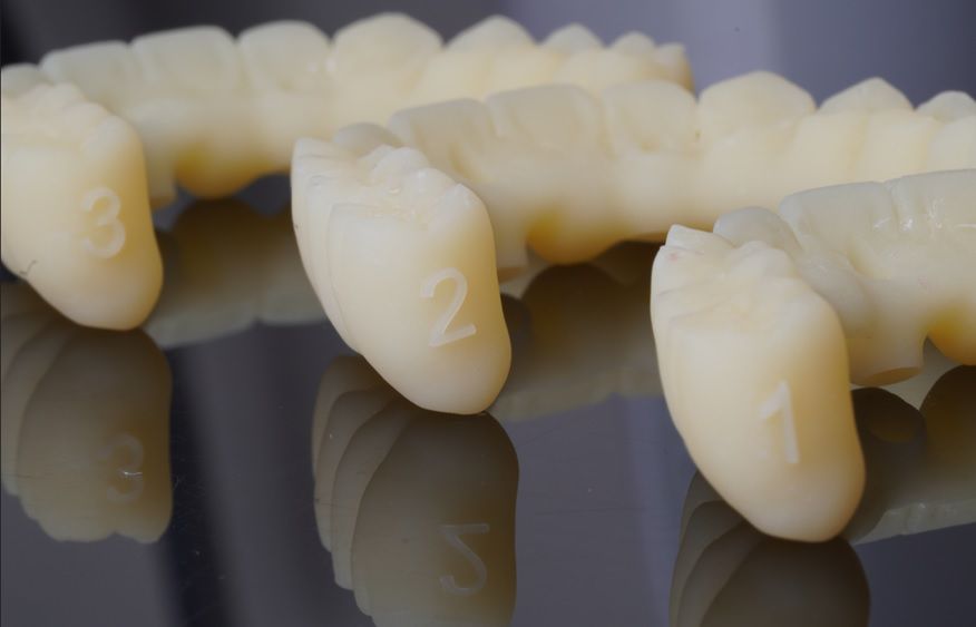
The three STL files obtained were sent to the digital laboratory for the design of the final proposal and the fabrication of three try-ins. Once the designs were approved, the laboratory provided three numbered try-ins (Figs. 16,17).

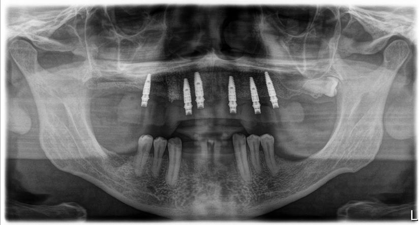
To assess the accuracy and clinical tolerance of the structures, a functional intraoral test of the try-ins was performed, for which the Variobases® were installed and subjected to a prosthetic screw passivity test. Finally, a standardized radiographic control was performed to verify the fit of the Variobase® to the SRA abutment.

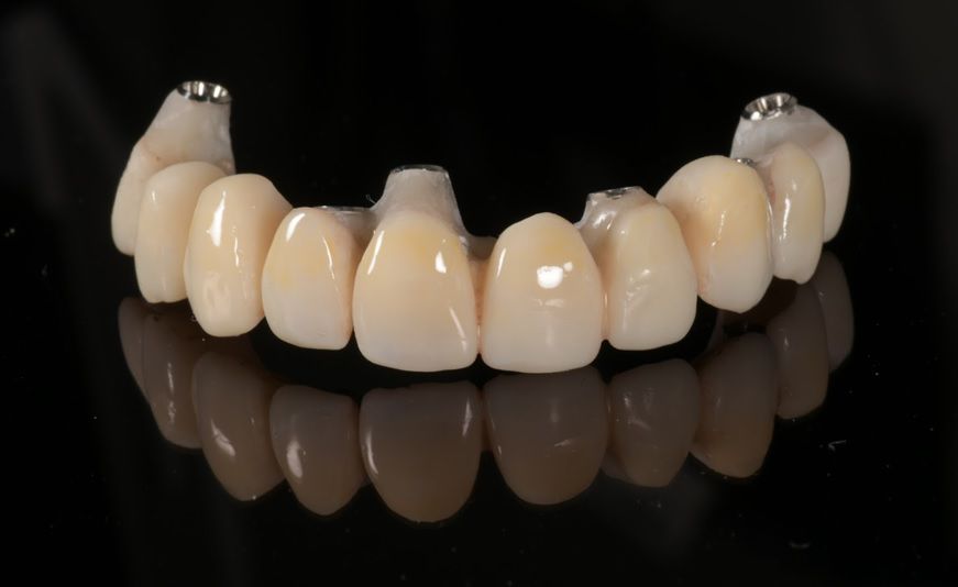
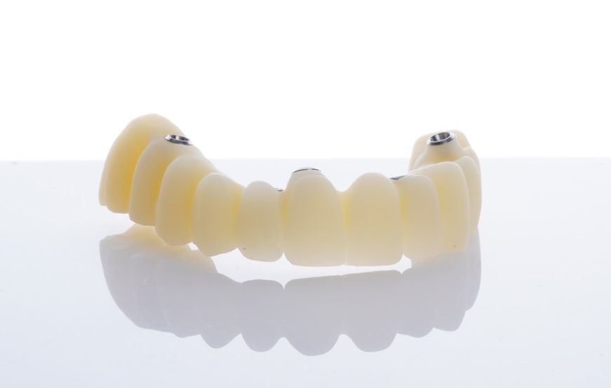
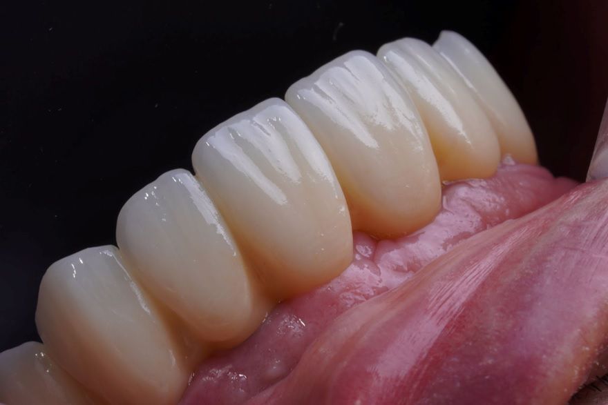
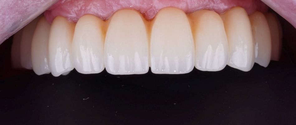
Once the correct adjustment of all the provisionals was confirmed, the decision was made to proceed with the one obtained using Methodology no. 1 (intraoral scanner and manually held provisionals). Esthetics and contours were refined to generate a new STL file for the subsequent fabrication of the definitive prosthesis. The monolithic zirconia prosthesis with titanium interfaces (Variobase®) was then cemented (Fig. 18).

Follow-up appointments were scheduled seven and thirty days after insertion. The use of intraoral irrigators was recommended for daily care, and a six-month appointment was indicated. At the end of the treatment, a maintenance schedule was established, with appointments every six months.

Treatment outcomes

No discrepancies were observed in the clinical or radiographic fit of the try-ins, according to the clinical evaluation by the operators. All three scan patterns achieved a correct and optimal clinical and radiographic fit of the try-ins (Figs. 19-23).

Author’s testimonial

Straumann® RevEX™ scanbody system is a true game changer in our workflow, allowing accurate, fast and simple transcription of implant position and soft tissue profiles into the digital environment of our lab and clinic. A must have in today's dentistry.

Related posts