#Full-Arch 17. Sep 2024

Total rehabilitation of a fully edentulous maxilla with immediate loading using Straumann® Pro Arch concept

A clinical case report by Tikarev Ravil

Full arch implant-supported restorations can restore function and aesthetics in edentulous patients. Advances in implant technology have significantly enhanced the ability to achieve predictable and long-lasting outcomes in these complex cases. The Straumann® BLX implant system, known for its surface technology and features that allow optimal primary stability, is particularly effective in immediate loading scenarios, making it a reliable option for full arch reconstructions.

Introduction

This case report focuses on the application of the Straumann® Pro Arch, which enables personalized treatment protocols for implant-borne fixed full-arch restorations. In this case, six BLX implants were placed in a fully edentulous patient with compromised oral function. The Straumann Pro Arch offers an efficient approach to full arch rehabilitation, combining the precision of guided surgery with the efficiency of immediate function. By integrating BLX implants into this treatment, clinicians can achieve stable and predictable outcomes, even in complex clinical situations.

The report outlines the clinical planning, surgical techniques, and prosthetic procedures involved in this case. It also highlights the rationale behind choosing the BLX implants, emphasizing how this approach can lead to predictable results in full arch restorations. Through this case, we aim to demonstrate the practical benefits of the Straumann® Pro Arch in delivering effective and lasting patient care.

Initial situation

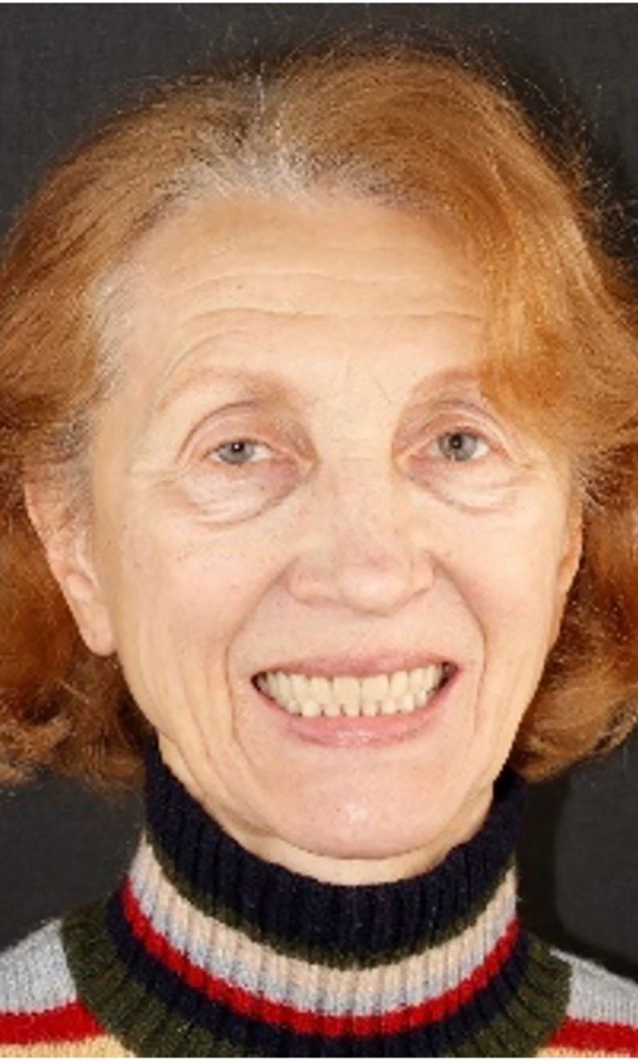
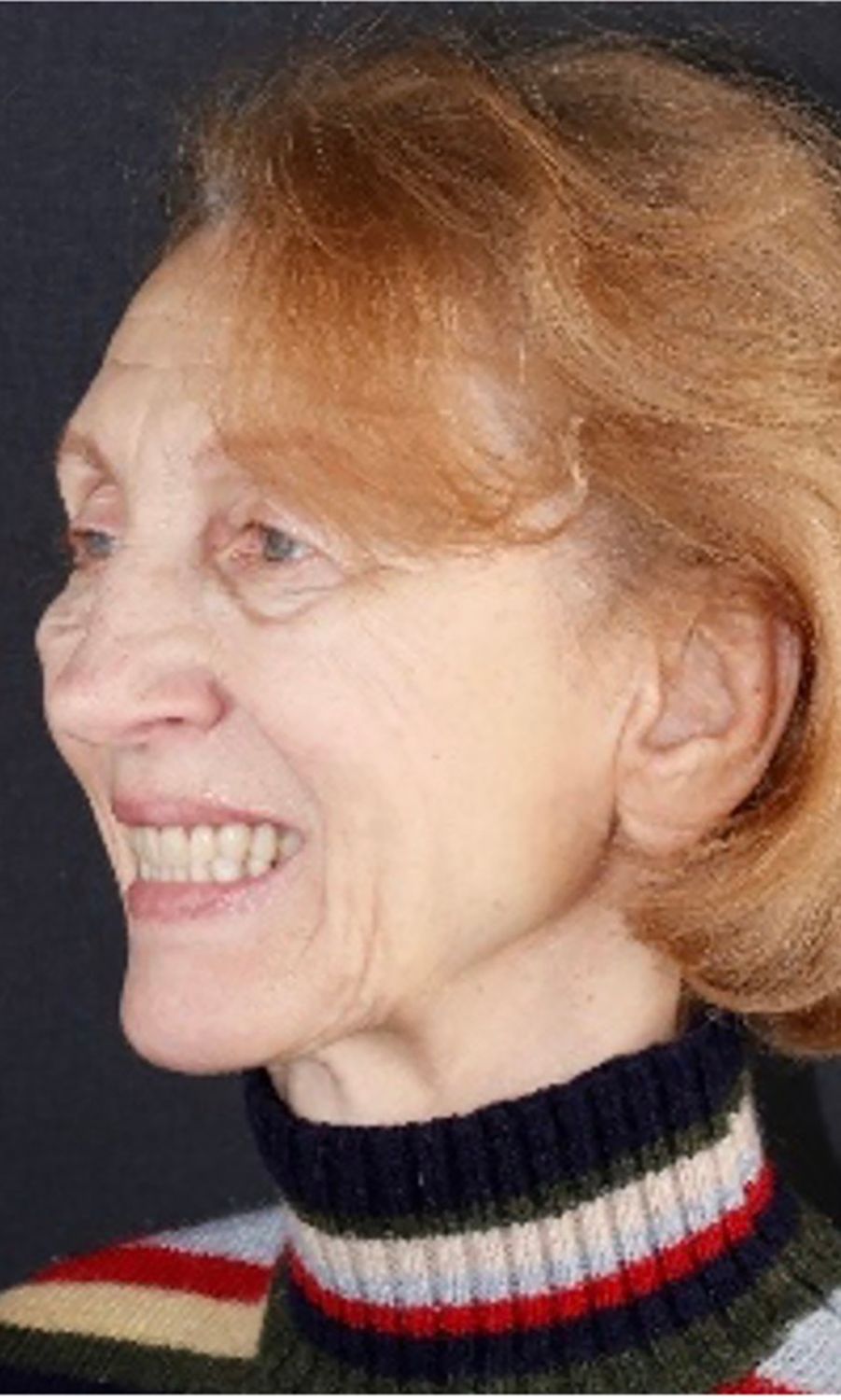
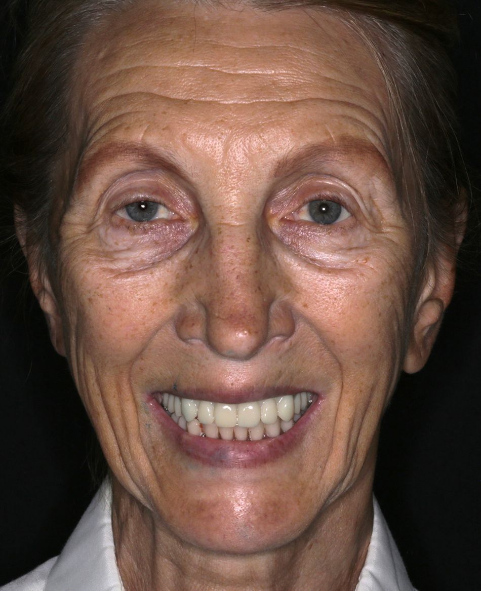
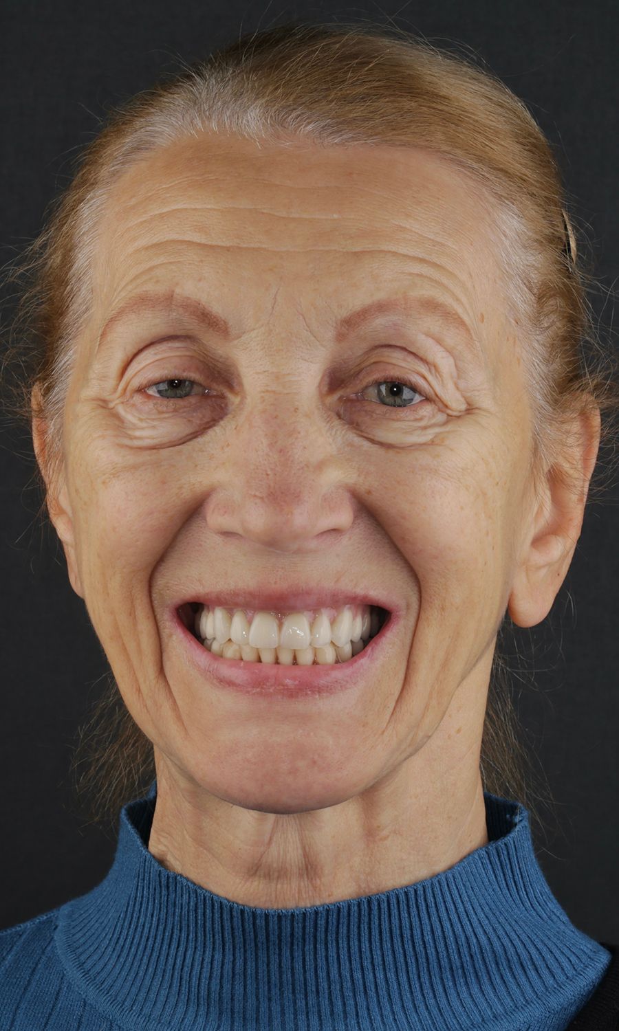
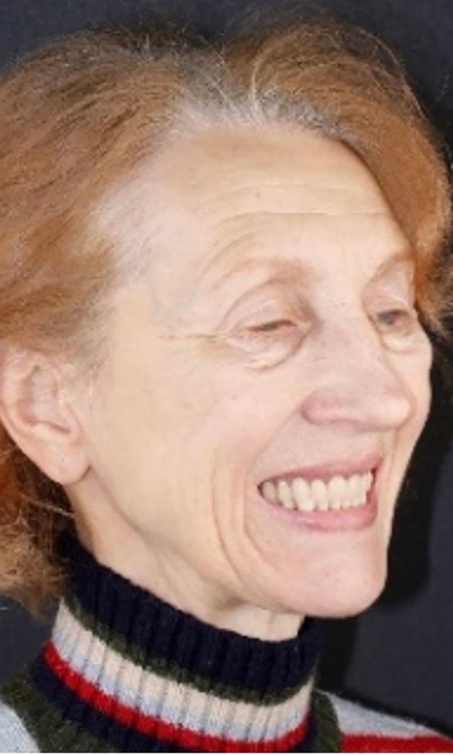
A 75-year-old female patient, classified as ASA I and a non-smoker, presented to our clinic with terminal dentition. She reported significant difficulty in chewing due to the instability of her upper denture and expressed a desire for a stable solution to rehabilitate her failing dentition and regain confidence in her ability to chew comfortably. Her primary request was to improve her current situation, enabling her to eat comfortably and confidently, thereby significantly enhancing her quality of life. During the extraoral examination, various parameters were evaluated. The patient demonstrated an adequate vertical dimension of occlusion and an appropriate incisal edge position. The position of her maxilla was found to be within normal ranges. The esthetics were acceptable. However, she had some difficulty with speech and, more significantly, with function, as the prosthesis was not stable (Figs. 1-3)

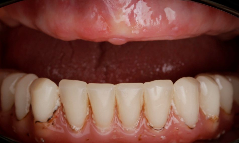
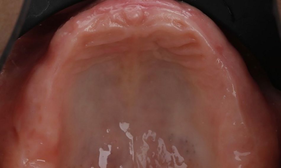
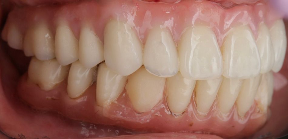
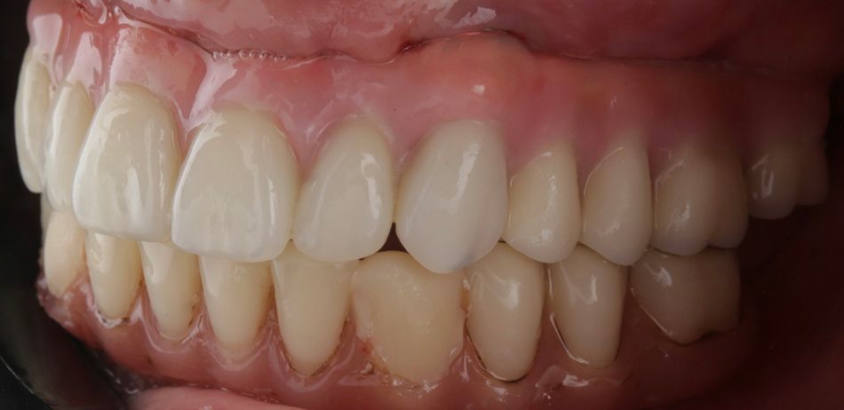
During the intraoral examination, the upper denture showed a lack of retention and stability. A midline discrepancy in relation to the facial midline was noted. Additionally, the patient presented with imbalanced occlusion when wearing the denture. After removing the upper denture, no intraoral pathology was detected (Figs. 4 & 5).

Treatment planning

After a thorough clinical evaluation, along with radiological (CBCT) and digital (intraoral) scanning, we proposed the following treatment plan: placement of six Straumann® BLX implants using guided surgery techniques, followed by the immediate loading of a temporary denture. Once adequate soft tissue healing was achieved, a fixed, non-removable full-arch prosthesis would be fabricated and fitted from an impression of the maxilla. This transformation will result in a fully stable and durable prosthesis, which will significantly improve the patient’s quality of life. The treatment workflow included:

  1. Placement of six Straumann® BLX implants using guided surgery techniques 
  2. Immediate loading of a temporary denture
  3. Fabrication and fitting of a fixed, non-removable full-arch prosthesis from an impression of the maxilla 

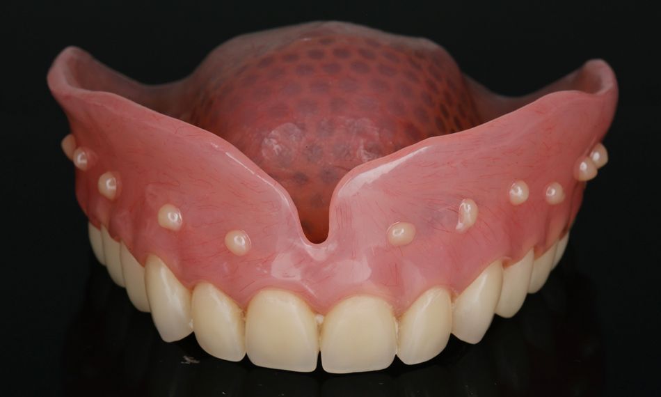
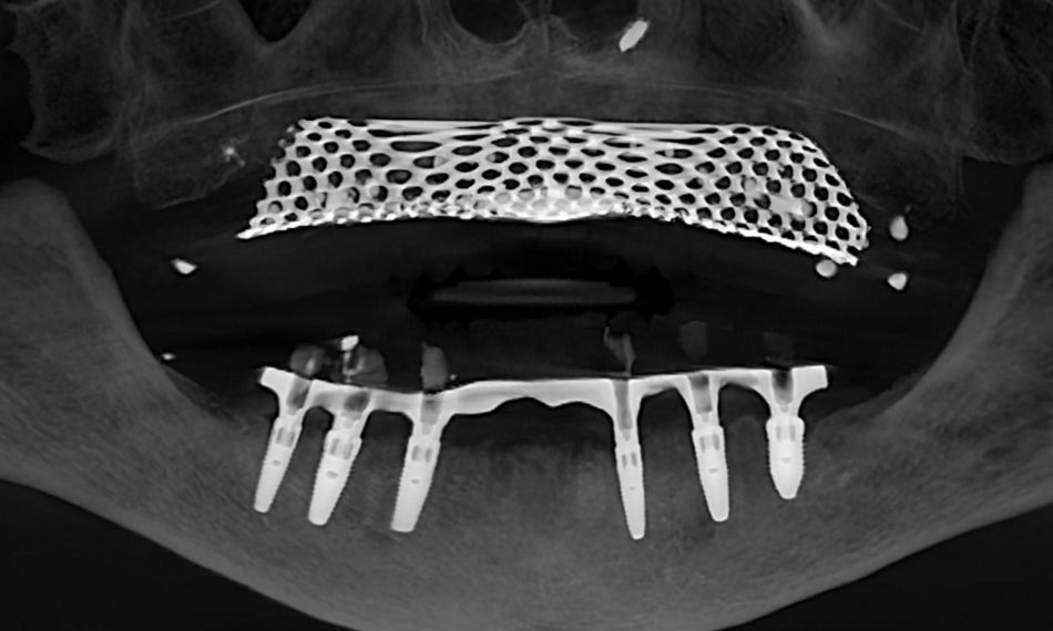
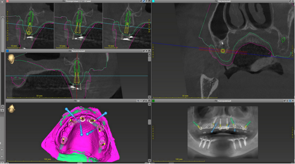
In order to plan a digitally guided surgery we performed CBCT with the full non-retained denture. We used light-cured material as markers to match the STL and DICOM files (Figs. 6 & 7). All planning was carried out in the coDiagnostiX® program.

Next, a complete denture was fabricated, incorporating a metal component on the palate. Unfortunately, this provided significant image artifacts when overlaying CBCT and DICOM files. In hindsight, it is sometimes necessary to make a new denture minus the metal part to prevent such artifacts.

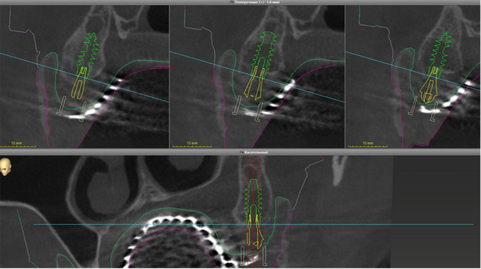
We planned to place the implants in areas with sufficient bone volume to ensure primary stability. Adequate alveolar bone width and height were present in the regions of both central incisors, first premolars, and second molars, allowing for ideal load distribution. To avoid intervention in the sinus area and minimize invasiveness, we decided to place four implants at an angled position (Figs. 8 & 9).

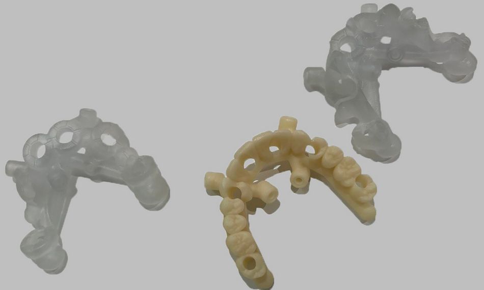
The template was planned with mucosa support to be fixed with pins on both the vestibular and palatine sides. The temporary prosthesis was made at the same time as the surgical template ensuring it could also be attached with pins (Fig. 10).

Surgical procedure

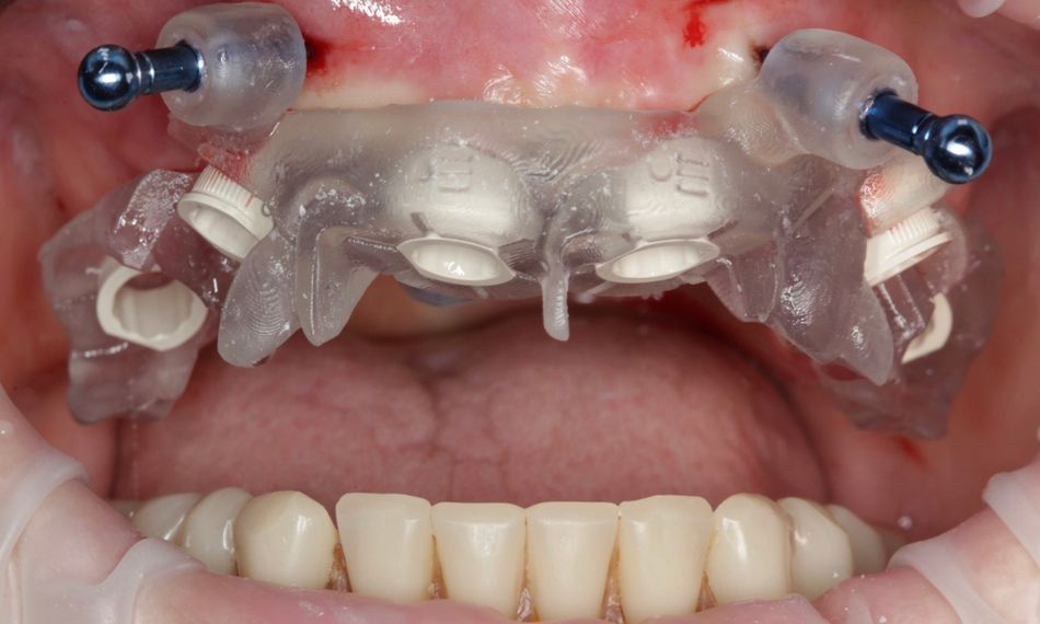
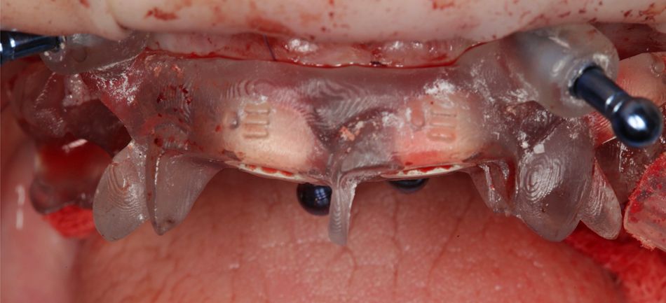
After administering general anesthesia with 1100 mg of propofol, the surgical template was securely attached to the patient using fixation pins. Sedation is often used to reduce discomfort, and we chose propofol for its effectiveness. Even with sedation, we ensured the patient’s comfort and satisfaction by administering a sufficient amount of local anesthetic. The procedure began with an incision in the gingiva, followed by the careful elevation of the flap to expose the underlying bone. The template was then meticulously positioned and secured, ensuring precise fitting for the guided surgery (Fig. 11). This step provided us with full surgical control and accuracy. Once the template was in place, we proceeded to shape the new gingival contour under the temporary prosthesis, ensuring optimal aesthetics and functionality for the patient’s immediate postoperative period (Fig. 12). Particular attention was given to ensure that the temporary prosthesis fit correctly with the newly contoured gingiva, promoting proper healing and comfort.

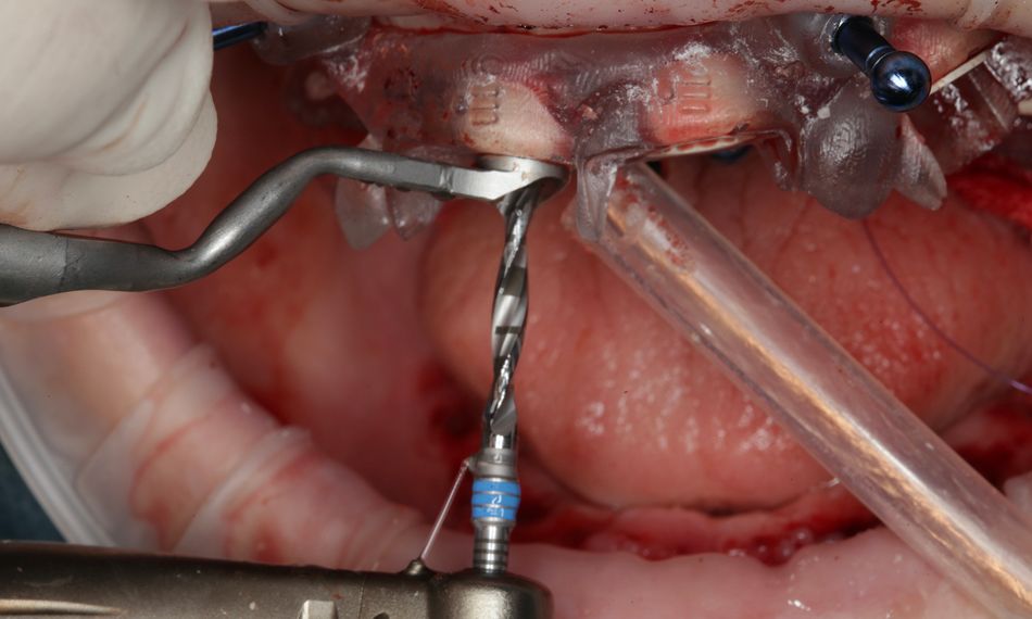
Next, we used the Straumann® VeloDrill™ System for guided surgery to ensure precise implant placement (Fig. 13). This advanced tool provided excellent navigational aid and significantly shortened the oral surgery chair time. We placed six Straumann® BLX implants in the positions of the central incisors, first premolars, and second molars. The Straumann® VeloDrill™ System allowed us to create exact osteotomies, ensuring each implant was inserted at the perfect depth and angle for optimal stability. Additionally, the Straumann® VeloDrill™ System minimizes heat generation and delivers high drilling stability. Continuous irrigation was maintained to protect the surrounding bone. Each implant was carefully torqued to achieve primary stability for the subsequent stages of the rehabilitation plan.

The implants used by location were the following:

• Implant #17:

o Straumann® BLX 4.5×12 mm.
o Primary stability: 45 N/cm.
o Straumann® Screw-retained Abutment RB 30°, 4.6 mm GH, 5.5 mm.

• Implant #14:

o Straumann® BLX 4.0×14 mm.
o Primary stability: 45 N/cm.
o Straumann® Screw-retained Abutment RB 30°, 4.6 mm GH, 4.5 mm.

• Implant #11:

o Straumann® BLX 4.0×12 mm.
o Primary stability: 50 N/cm.
o Straumann® Screw-retained Abutment RB, 4.6 mm GH, 3.5 mm.

• Implant #21:

o Straumann® BLX 4.0×12 mm.
o Primary stability: 45 N/cm.
o Straumann® Screw-retained Abutment RB, 4.6 mm, 3.5 mm.

• Implant #24:

o Straumann® BLX 4.5×14 mm.
o Primary stability: 50 N/cm.
o Straumann® Screw-retained Abutment RB 30°, 4.6 mm, 4.5 mm.

• Implant #27:

o Straumann® BLX 4.5×12 mm.
o Primary stability: 25 N/cm.
o Coverscrew.

Straumann® XenoGraft® 0.25 was used to perform horizontal bone augmentation around implants #11 and #21 to improve the bone volume and provide a stable base for the implants. The graft material, which is well-known for its capacity to support bone regeneration, was carefully placed. Following the bone augmentation, the flap was closed using Monosyn 5/0 material, which is a sterile synthetic absorbable monofilament. This choice of suture was made for its reliable strength and minimal tissue irritation, facilitating effective wound closure and promoting optimal healing.

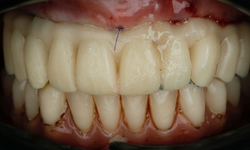
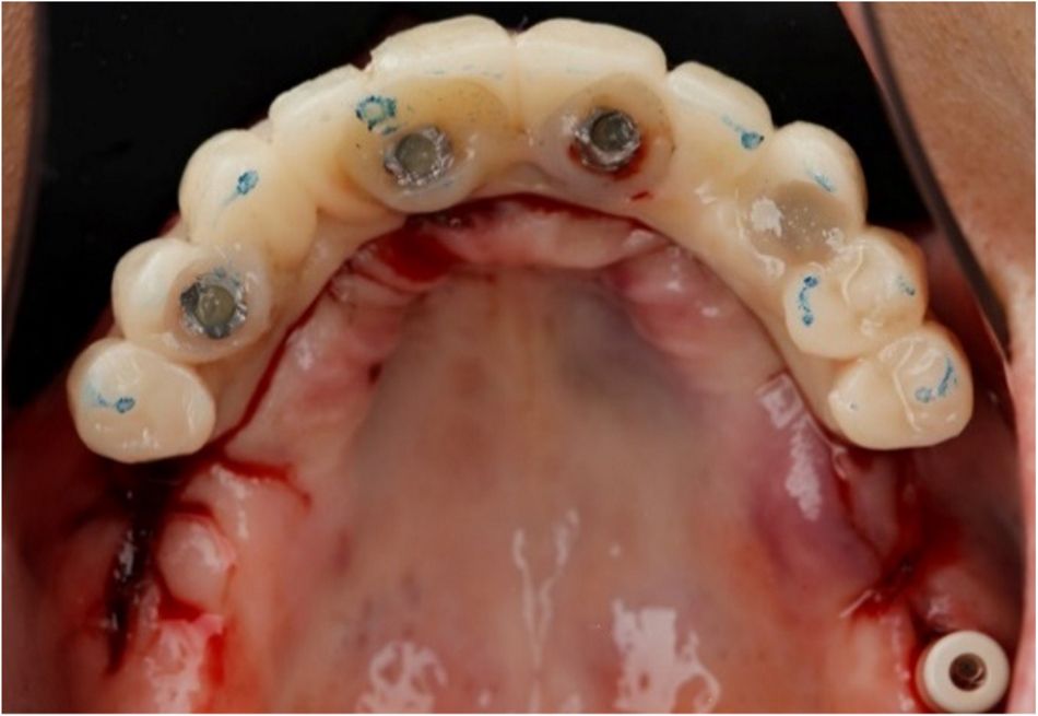
Once the implants were properly positioned, six temporary copings were screwed to the Straumann® screw-retained abutments (SRA) to simplify the fabrication of the multi-unit temporary screw-retained prosthesis, which was designed to offer immediate functional and aesthetic support. These titanium copings were fixed in place after carefully suturing the gingiva to ensure proper tissue adaptation and healing. During this step, aspects such as occlusion, esthetics, phonetics, and vertical dimension were carefully checked (Figs. 14 & 15). The temporary screw-retained prosthesis was then sent to the laboratory for polishing, contour refinement, and final adjustments. Upon completion, the prosthesis was attached to the implants using a light-curing material, ensuring a stable fit and optimal functional and esthetic outcomes during the healing phase (Fig. 16).

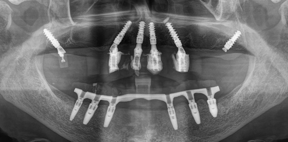
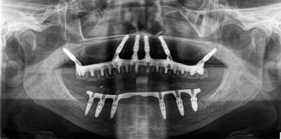
Finally, an X-ray was taken to verify the correct positioning of the implants and the proper fit of the prosthesis (Fig. 17).

Prosthetic procedure

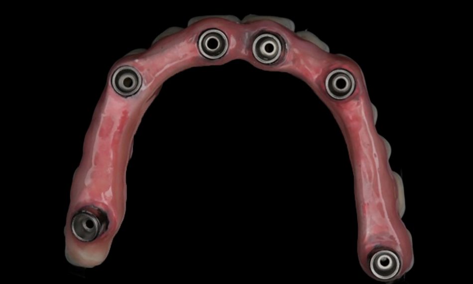
Four months later, after the implants had been placed and osseointegration along with soft tissue healing were completed, an impression of the patient’s upper jaw was taken. This impression was used to create a prototype for the final restoration (Fig. 18).

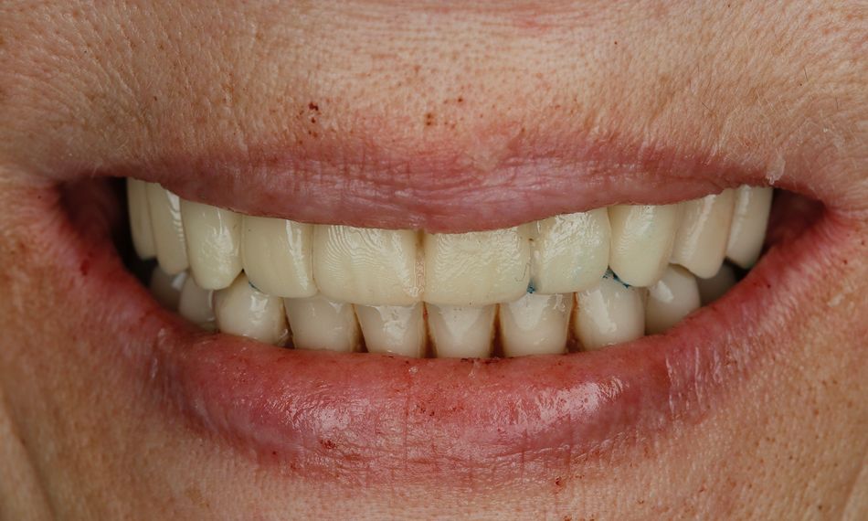
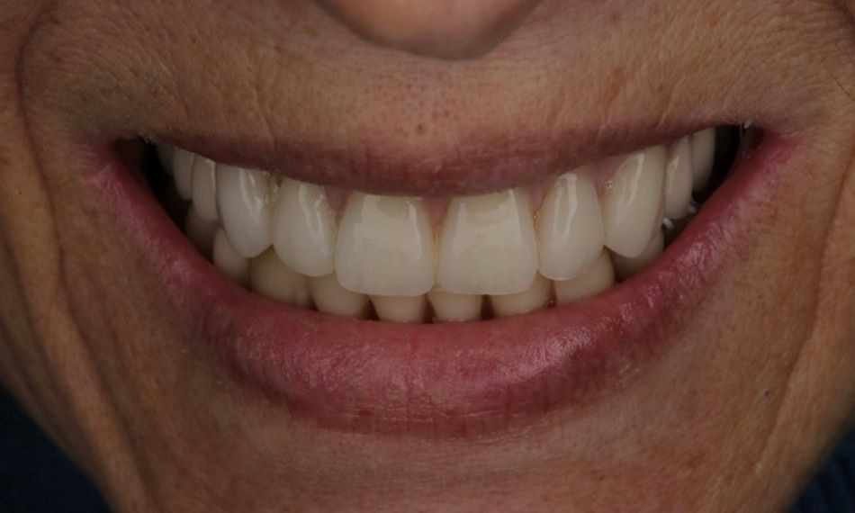
Once the patient approved the prototype, the final restoration was delivered. We then rechecked all aspects, including occlusion, function, and aesthetics, to ensure everything was perfect (Figs. 19 - 22). To enhance the prosthetic's durability, the restoration included the addition of a titanium bar and was secured with occlusion screws with a torque of 15 N/cm (Fig. 23). Articulation paper was used to identify further occlusion to 50 micrometers.

A final radiograph taken at the time of definitive prosthesis delivery showed no signs of bone loss, thereby confirming the successful outcome of the restoration (Fig. 24).

The patient received detailed instructions on maintaining proper hygiene and was provided with a comprehensive care plan. This plan included scheduling follow-up appointments for regular monitoring and any necessary adjustments.

Treatment outcomes

The patient was extremely pleased with the screw-retained prosthesis, which allowed her to eat and chew comfortably and without any concerns. This significantly enhanced her quality of life, aligning perfectly with the Straumann® Pro Arch concept’s goal. The use of surgical navigation made the procedure less invasive and more predictable, simplifying the process for both the patient and the surgeon.