#Full-Arch 27. Sep 2023

Maxillary full-arch, implant-supported rehabilitation with a digital workflow: 2-year follow-up

A clinical case report by Li Xiaofeng and Li Shuguang, China

Complete edentulism is an irreversible condition explained as the known marker of disease burden for oral health where an oral cavity is without any teeth. (1) Edentulism significantly influences both oral and overall health, as well as the quality of life.(2) In modern times, implant-supported full-arch rehabilitations have emerged as a reliable and foreseeable treatment option for edentulism cases.(3)

Introduction

New developments, such as virtual-guided technologies, offer an optimal approach for implant placement, aligning with the accurate prosthetic position and minimizing both intraoperative trauma and procedure duration. 4

The following case report demonstrates the successful management of a 70-year-old patient experiencing chewing difficulties. Through a maxillary full-arch, implant-supported rehabilitation with six Straumann® BLT implants facilitated with a digital workflow, we improved her quality of life and fulfilled her expectations.

Initial situation

A 70-year-old healthy female with no medication, allergies, or smoking habits came to our clinic seeking a resolution for her difficulties with chewing. Additionally, she reported a history of persistent dental and oral malodor issues. The patient expressed a desire to “go back in time” and be able to eat like before and recover her quality of life and confidence.

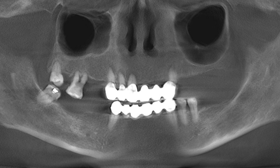
The extraoral examination showed a low smile line. On the other hand, the intraoral examination depicted a bridge from #13-#23 in hopeless conditions. The teeth of the upper jaw, with the exception of #18, were mobile. Generalized gingival inflammation, bleeding on probing and dental caries were also present (Fig. 1). In addition, the patient exhibited a removable prosthesis in the upper jaw that was poorly adapted and showed signs of deterioration. The radiographic examination exposed severe bone loss on the posterior side of the 2nd quadrant. Furthermore, images were also compatible with caries and apical lesions on maxillary teeth (Fig. 2).

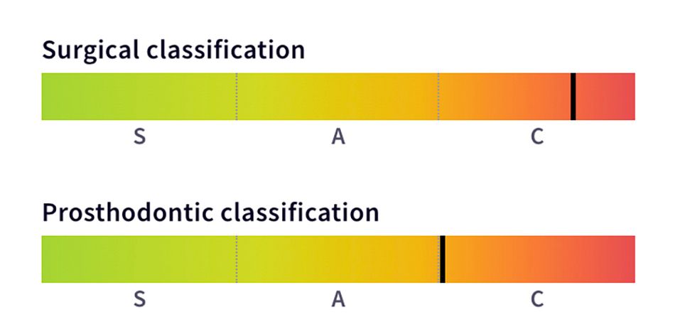
After conducting both radiographic and clinical assessments, the patient was categorized as having a complex condition according to both the surgical and prosthodontic SAC classifications. This classification system assists in evaluating the level of challenge and potential complications linked to implant-related restoration (Fig. 3).

Treatment planning

Following an extensive conversation about the different available treatment alternatives with the patient, a joint decision was reached, and it was concluded that the chosen approach would involve a digital workflow, immediate implant placement, and a maxillary full-arch, implant-supported restoration.

The treatment workflow included:

  1. Digital implant placement planning using coDiagnostiX®.
  2. Production of the printed surgical guide and resin models.
  3. Extraction of hopeless teeth in the upper jaw.
  4. Fixation of the guide and immediate implant placement of six Straumann® BLT implants in the maxilla.
  5. Filling of the gaps with a Straumann® XenoGraft.
  6. Temporary screw-retained prosthesis delivery.
  7. Final screw-retained prosthesis delivery 12 weeks after surgery.
  8. Periodontal supportive therapy (every 3-4 months). 

Using coDiagnostiX® for digital planning of implant placement proved to be of paramount significance in safeguarding the adjacent anatomical structures and achieving consistent and reliable outcomes. Additionally, this software facilitated the creation of a highly precise surgical guide tailored to our patient's needs. Upon finalizing the design, the next step involved producing the surgical guides through 3D printing and crafting resin models.


A Center of Dental Education (CoDE) is part of a group of independent dental centers all over the world that offer excellence in oral healthcare by providing the most advanced treatment procedures based on the best available literature and the latest technology. CoDEs are where science meets practice in a real-world clinical environment.


Surgical procedure

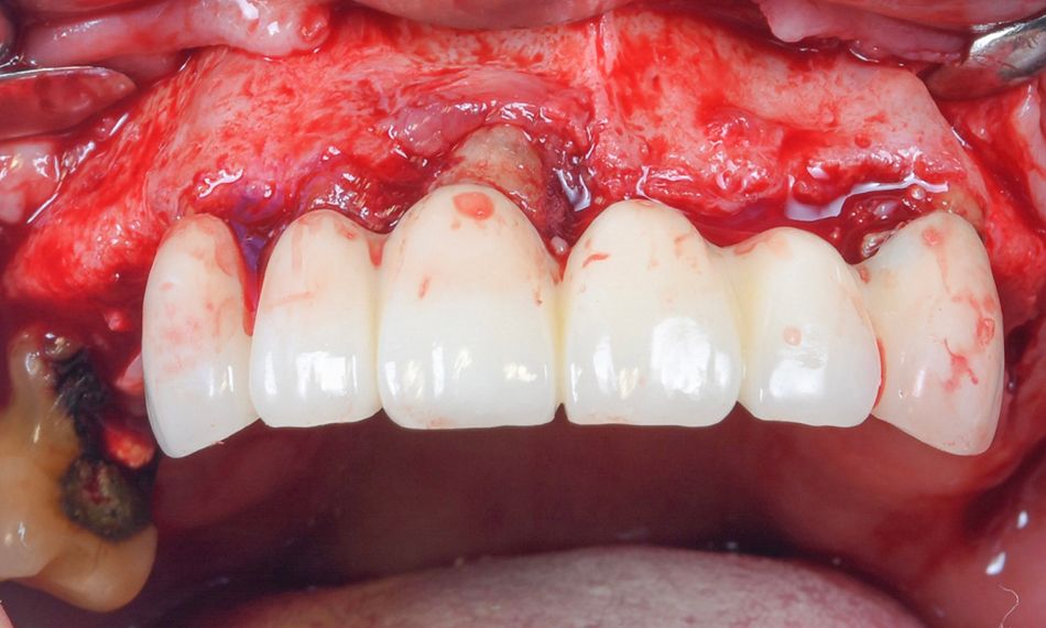
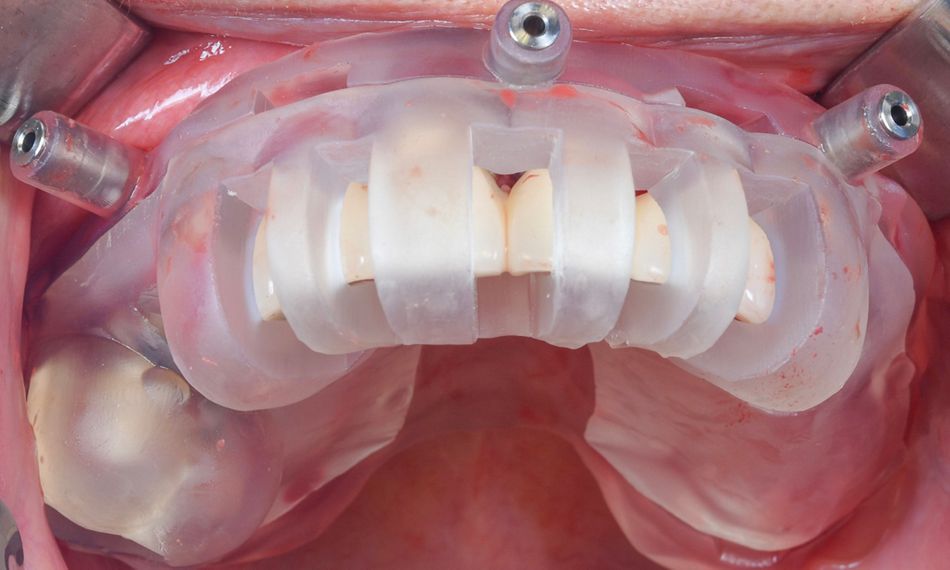
Prior to the surgical procedure, a careful assessment was conducted to ensure the precise fitting of the printed guide. Subsequently, local anesthesia using 2% lidocaine with 1:100k epinephrine was administered. An open flap access was performed with mid-crestal and intrasulcular incisions (Fig. 4). Next, the bone reduction guide was placed, and the fixation pins were drilled and securely inserted (Fig. 5).

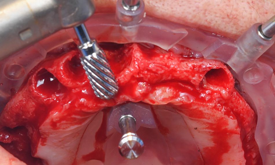
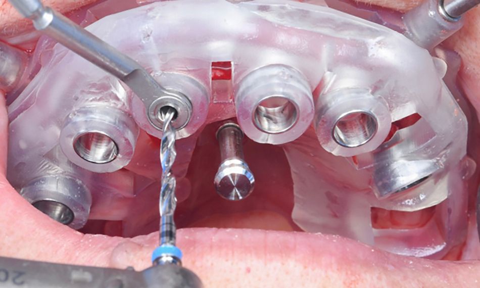
The bridge was removed, and the teeth were atraumatically extracted. All extraction sockets underwent a meticulous curettage. Following the dental extractions, a reduction of the vertical ridge was carried out (Fig. 6). Subsequently, the osteotomy guide was positioned to initiate the drilling procedure for the immediate implant placement (Fig. 7).

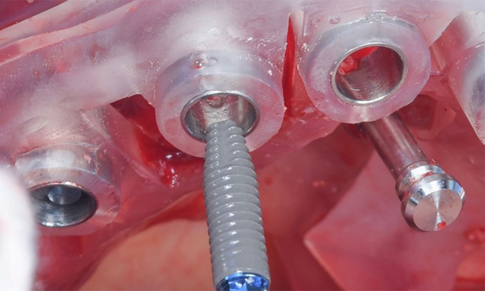
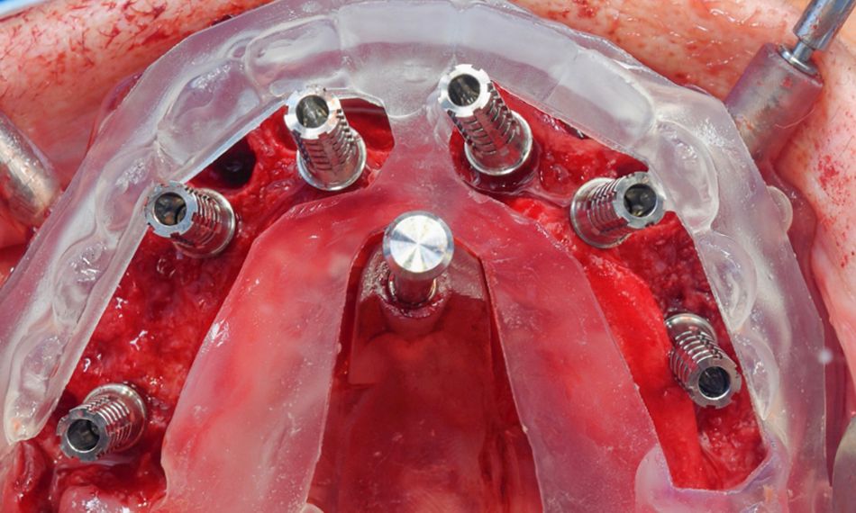
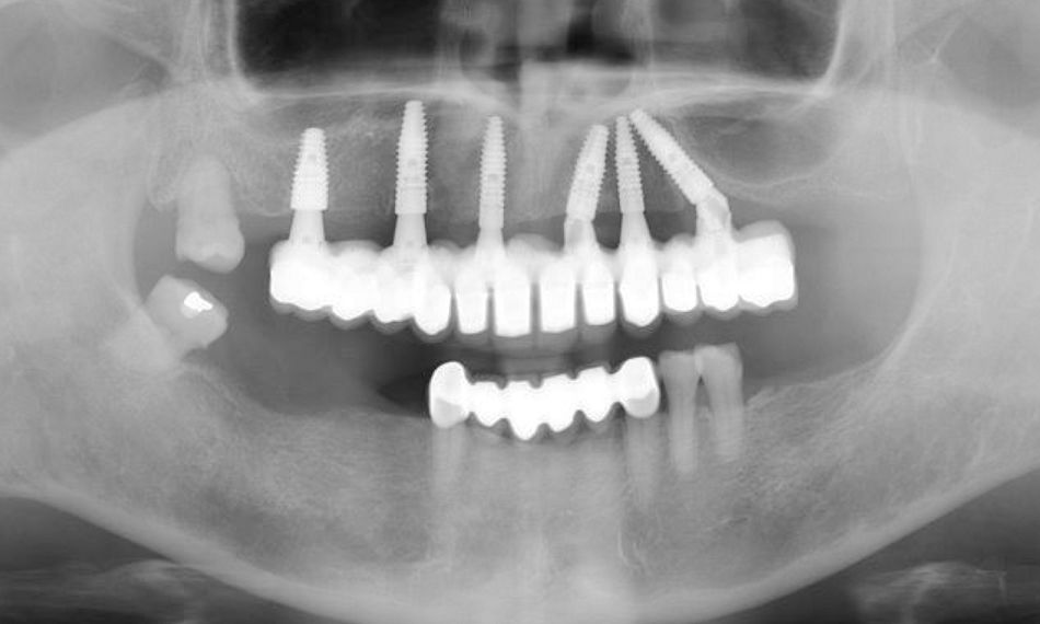
Six Straumann® BLT implants made from the material Roxolid® and with the SLActive® surface (16,4.8*10 mm; 14, 4.1*14 mm; 12, 3.3*14 mm; 21, 4.1*12 mm; 23, 3.3*12 mm; 26, 4.1*14 mm ) were placed in the maxilla (Fig. 8). The gaps were filled with Straumann® XenoGraft, and six temporary titanium copings for screw-retained abutment were subsequently placed (Fig. 9).

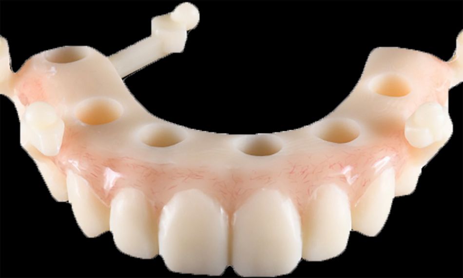
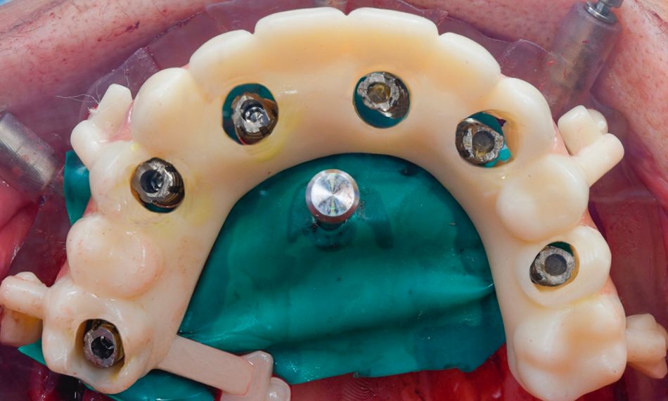
Full-arch maxillary impressions were taken, employing appropriate transfers for an open-tray approach using a polyether impression material (Impregum, 3M ESPE). After a few hours, a rubber dam was placed around the temporary copings to protect the fresh surgical sites. The previously prepared temporary prosthesis produced from resin-based provisional material was then affixed in place (Figs. 10-12).

The patient was prescribed painkillers and antibiotics and given instructions for postoperative oral care, which included rinsing the oral cavity twice a day for a week with a 0.2% chlorhexidine gluconate mouthwash and cleaning the temporary restorations with a very soft toothbrush.

The sutures were removed 12 days after the surgical procedure.

Prosthetic procedure

After 12 weeks, the delivery of the final screw-retained restoration was planned. During this time, the implant sites underwent successful healing, and osseointegration was accomplished.

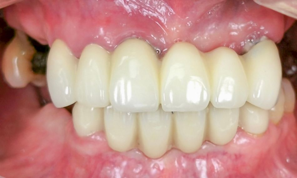
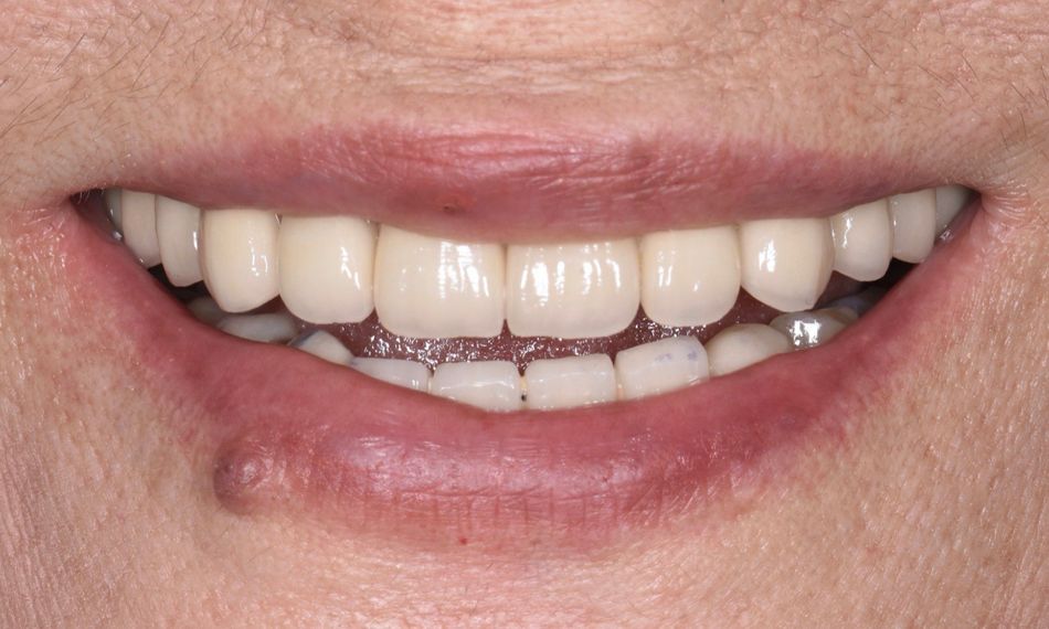
An open-tray impression was taken, leading to the acquisition of final cast models. These models were then utilized to create the final implant-supported full-arch maxillary rehabilitation. A comprehensive assessment was conducted on the full-arch prosthesis to ensure precise and passive fitting during the clinical evaluation. Furthermore, functionality, phonetics, occlusion, and esthetics were meticulously verified (Fig. 13). The screw access holes were filled using Filtek Supreme resin (3M ESPE).

Oral hygiene instructions were given, and a periodontal supportive therapy was coordinated for every 3-4 months.

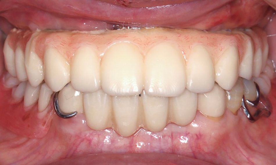
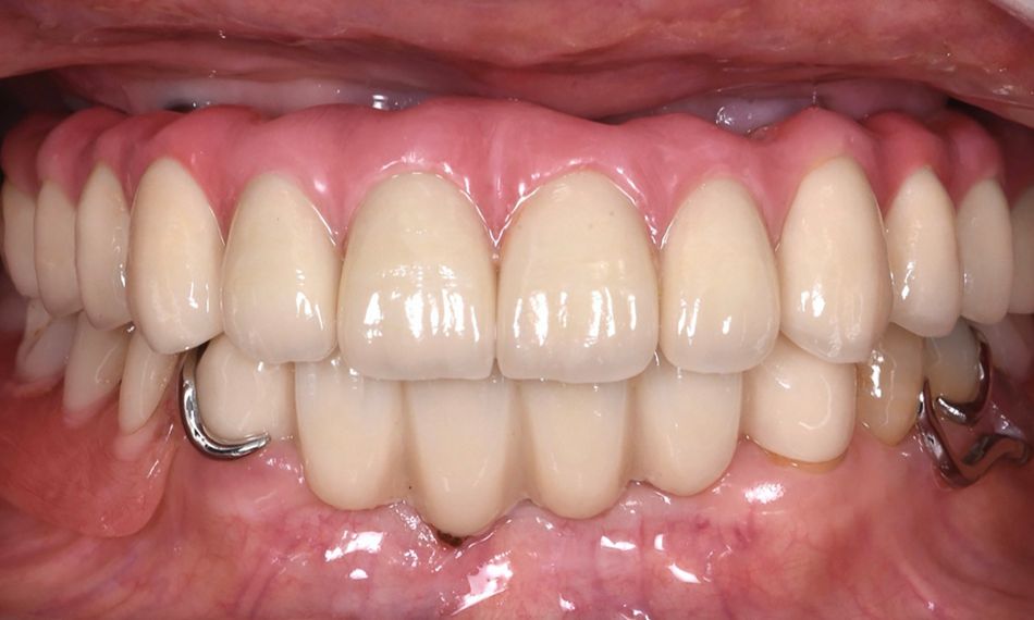
After two years, a clinical and radiographic control of the rehabilitation on implants was carried out (Figs. 14,15). The soft and hard tissues surrounding the implant exhibited favorable conditions. Furthermore, the patient has gained a deeper understanding of her oral health and has acquired knowledge on how to maintain proper oral care.


Treatment outcomes

The patient expressed great satisfaction with the achieved outcomes. She could now enjoy eating without any concerns. Similarly positive outcomes were achieved across functional, biological, and esthetic aspects.

Author’s testimonial

Immediate implant placement after tooth extraction has become a common surgical approach in clinical practice. The outcome of this procedure is as predictable as implant placement in healed sites. In patients with periodontal compromise and hopeless teeth, they are an excellent alternative for rehabilitation, as long as adequate access for peri-implant hygiene is ensured.

References:

  1. Cunha-Cruz J, Hujoel PP, Nadanovsky PA. Secular trends in socio-economic disparities in edentulism: USA, 1972–2001. J Dent Res. 2007;86:131–6.
  2. Abdunabi A, Morris M, Nader SA, Souza RF. Impact of immediately loaded implant-supported maxillary full-arch dental prostheses: a systematic review. J Appl Oral Sci. 019:12;27:e20180600.
  3. Pera P., Menini M., Pesce P., Bevilacqua M., Pera F., Tealdo T. Immediate Versus Delayed Loading of Dental Implants Supporting Fixed Full-Arch Maxillary Prostheses: A 10-year Follow-up Report. Int. J. Prosthodont. 2019;32:27–31.
  4. Ashurko I, Trofimov A, Tarasenko S, Mekhtieva S. Full-Mouth Screw-Retained Implant-Supported Rehabilitation with Multiunit Abutments Using Virtual Guided Surgery and Digital Prosthetics Protocol. Case Rep Dent.;2020:3585169.