#Edentulous 18. Feb 2021

Precise prosthetic planning & implant placement: treatment of a complex full-arch case using guided surgery and immediate loading

A clinical case report by Sergio Piano, Italy

Oral health plays an important role in quality of life and even though preventive dentistry has evolved, edentulism remains a prominent public health problem that can affect function, esthetics, and psycho- social aspects of the human being. Thus, the option of replacing teeth with implants plays an important role in patient wellbeing.

Introduction

Technological progress, including the proliferation of internet access worldwide, has given patients access to a wealth of information about their oral health and has raised awareness of the range of available treatments, materials and techniques. As a result, patients’ expectations and behaviors have changed and an ever-increasing number are requesting less invasive, affordable, efficient, and immediate solutions.

The Straumann® Pro Arch Concept enables personalized treatment protocols with various treatment options to address specific indications and clinical scenarios. This concept covers all stages of an immediate full arch treatment and includes the use of implants specifically designed for immediate procedures, with a flexible prosthetic portfolio that considers patients’ esthetic expectations and financial resources.

The following case report describes a successful functional and esthetic prosthetic full-arch implant rehabilitation of a patient with a hopeless maxillary dentition and decreased quality of life due to impaired masticatory function and poor esthetics. The Straumann® Pro Arch Concept with Straumann® Bone Level Tapered (BLT) implants and Straumann® Screw Retained abutments (SRA) allowed us to address our patient’s chief complaints by providing an efficient and predictable solution. The immediate treatment workflow involved 4 steps; 1) accurately creating a pre- operative dental model; 2) surgical and prosthetic planning with coDiagnostiX® software; 3) placing the dental implants with computer-guided and flapless technique; and 4) finally performing an immediate loading procedure.

This clinical outcome was achieved in a single day and with no complications. The patient was highly satisfied with the positive impact the treatment had on her quality of life due to significantly improved esthetics and recovery of masticatory function.

Initial situation

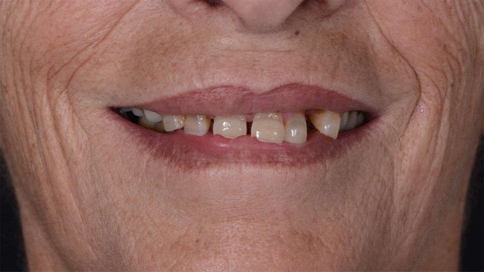
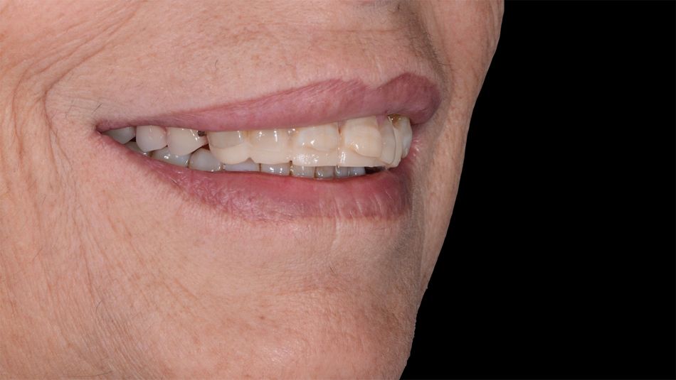
A systematically healthy 58-year-old female patient presented to our office with the chief complaint of esthetic dissatisfaction and impaired masticatory function due to an unstable removable partial denture (Fig. 1).

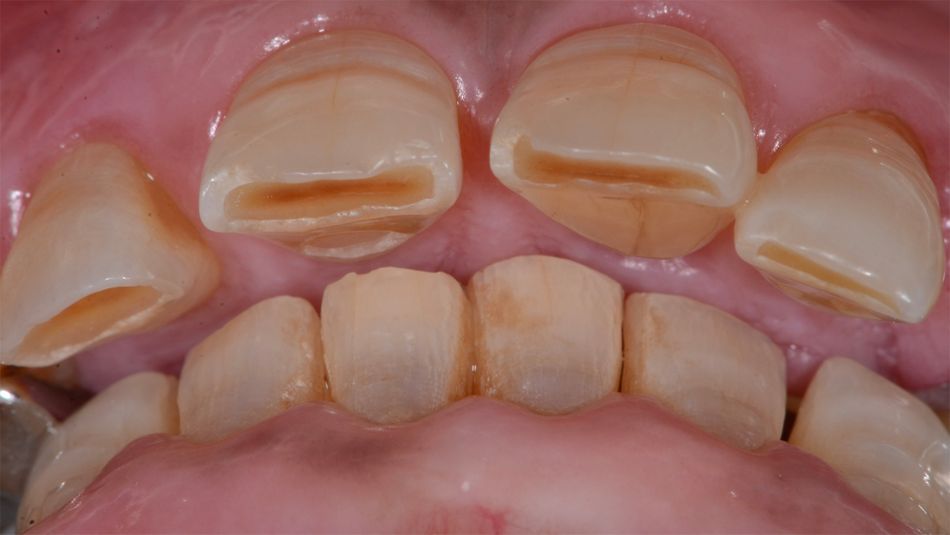
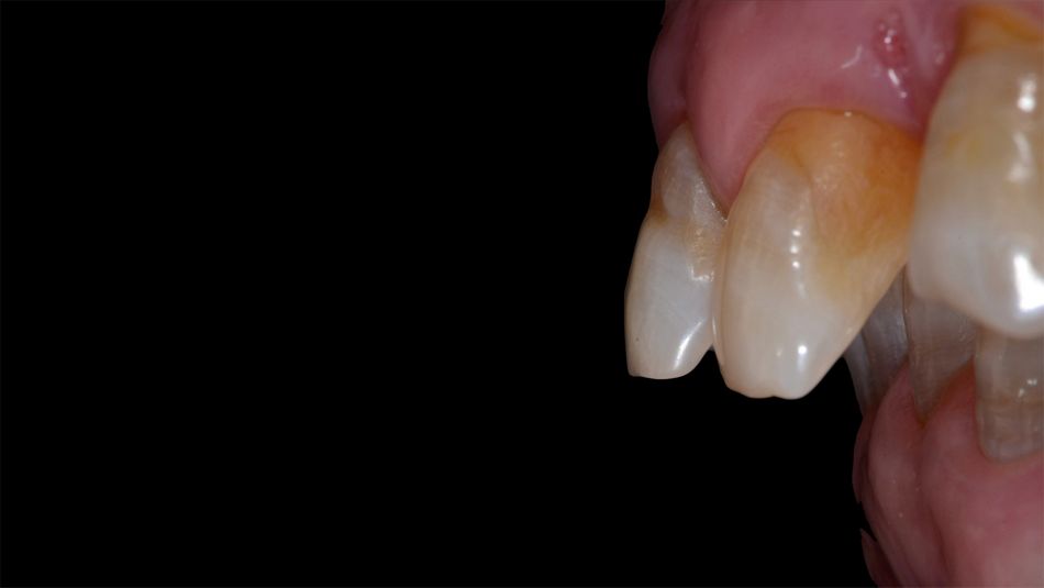
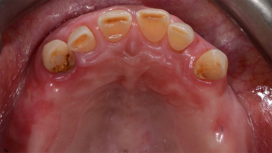
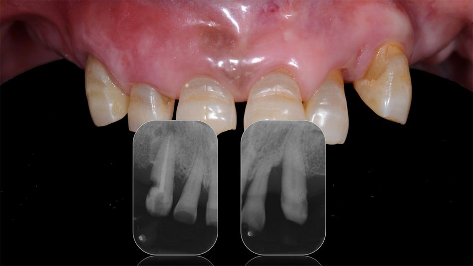
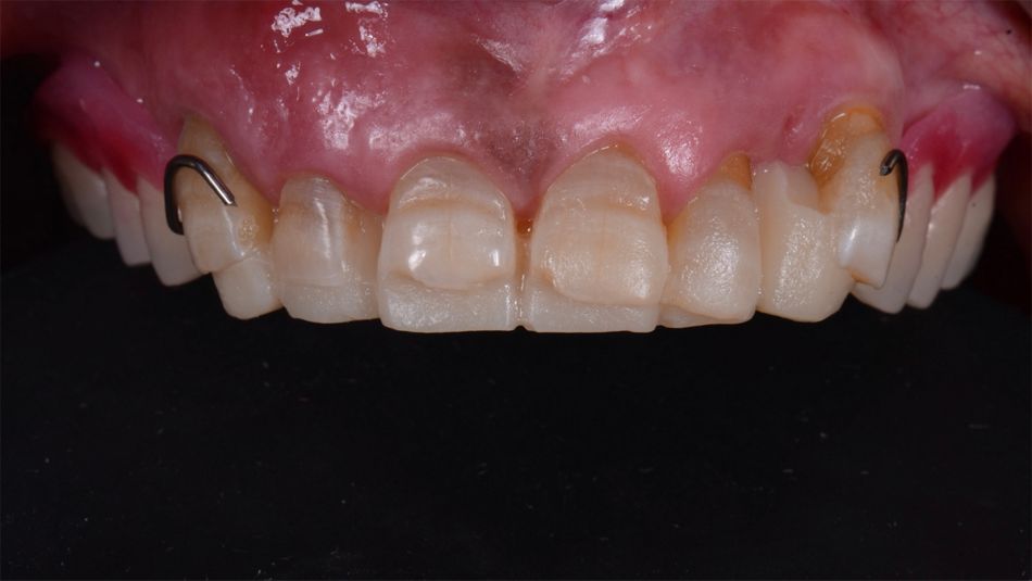
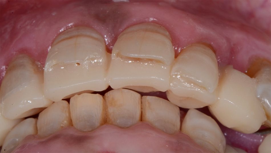
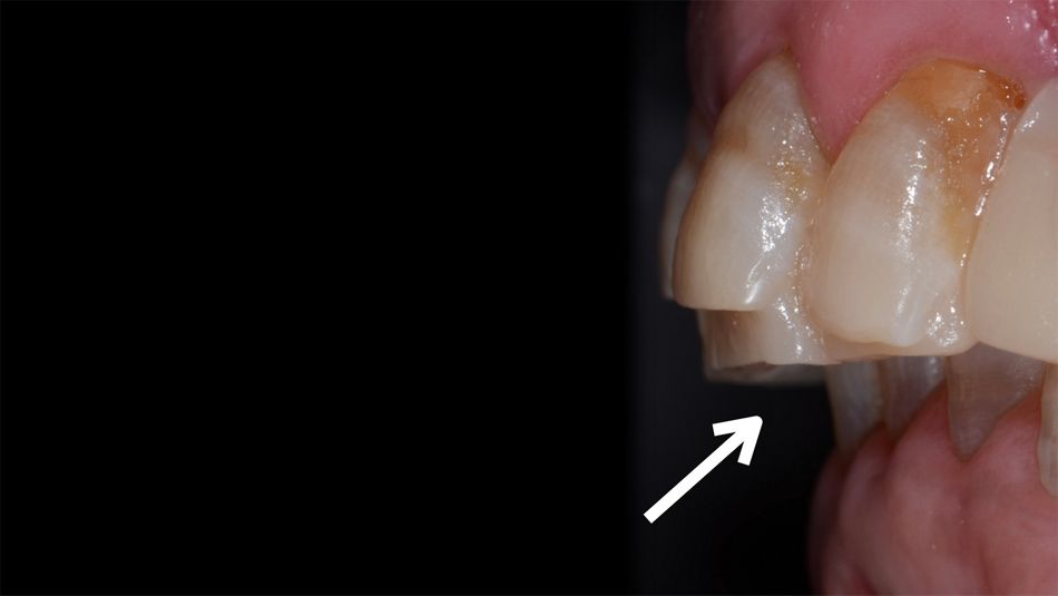
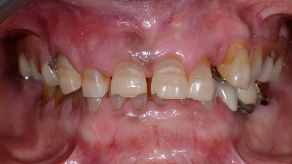
The initial comprehensive clinical evaluations revealed partial edentulism, a deep overbite with the lower incisors impinging upon the palatal gingiva and increased overjet. The upper incisors were buccally tilted and presented tooth wear and mobility. Moreover, there was interference between the lower lip and the anterior teeth in the smile position (Figs. 2-5).
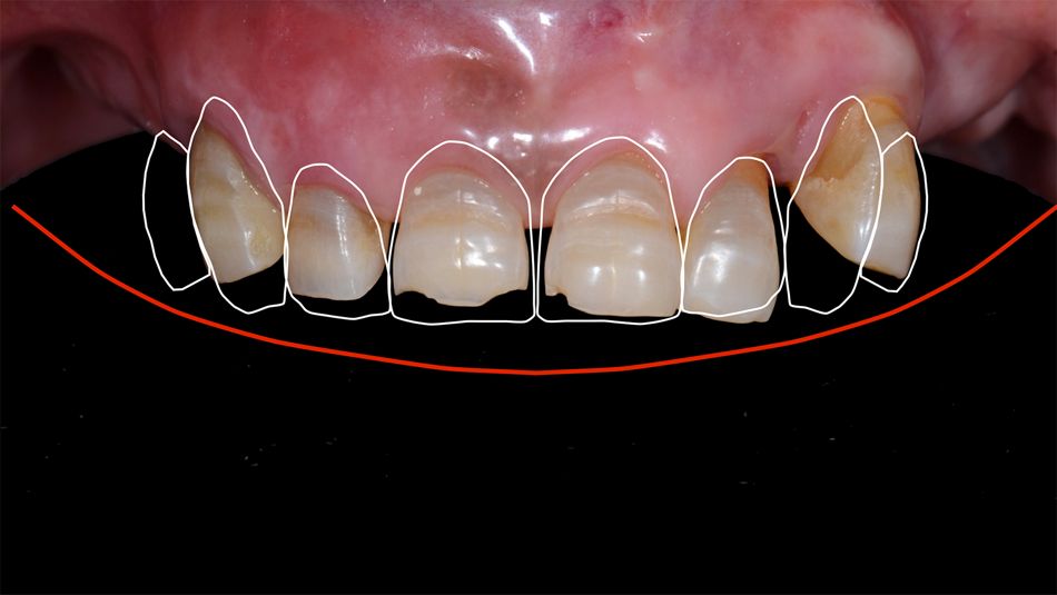
Intraoral periapical X-rays showed approximately 50–75% alveolar bone loss. In addition, the patient had an uneven smile line (Fig. 6,7).

Treatment planning

The benefits, risks, and alternative treatment options were discussed with the patient and a decision was then reached in partnership with the patient. This is a fundamental stage in treatment planning to ensure that the patient's needs and wishes are met and to achieve a successful outcome.

The treatment workflow included the immediate placement of implants into the extraction sockets and immediate loading using computer-guided surgery and a flapless approach.

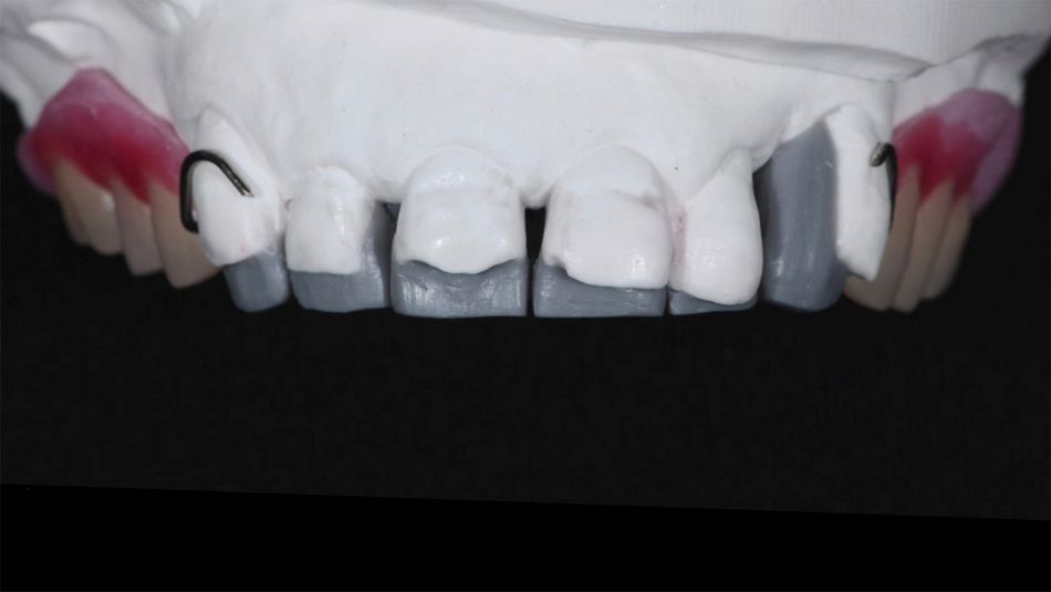
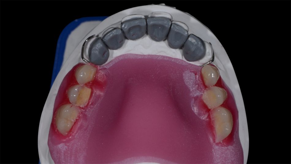
A suitable new removable partial denture was planned as a starting point. Following the clinical documentation, a digital smile design (DSD) preview was created. The dental technician then made a wax-up of the frontal teeth and a model of the posterior area, thereby creating a new dental arrangement. This new model increased the vertical dimension to define an optimal vertical position for the front teeth (Figs. 8,9).

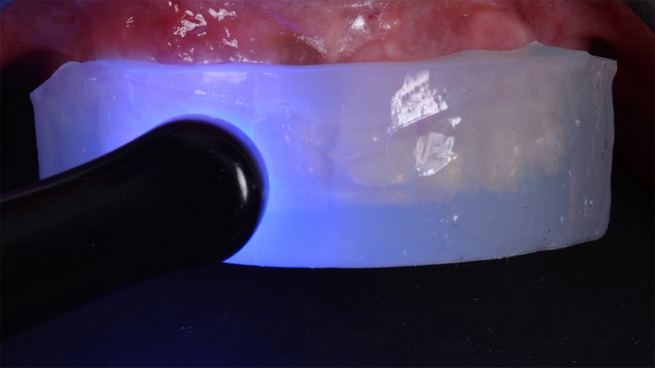
The next step was to transfer this data to the patient's mouth to verify its accuracy. Using a silicone mask created based on the wax-up and filled with flowable composite, a mock-up of the front teeth was made (Fig. 10). A set-up of the posterior teeth was added to the mock-up to complete the previsualization of the proposed new smile (Fig. 11).

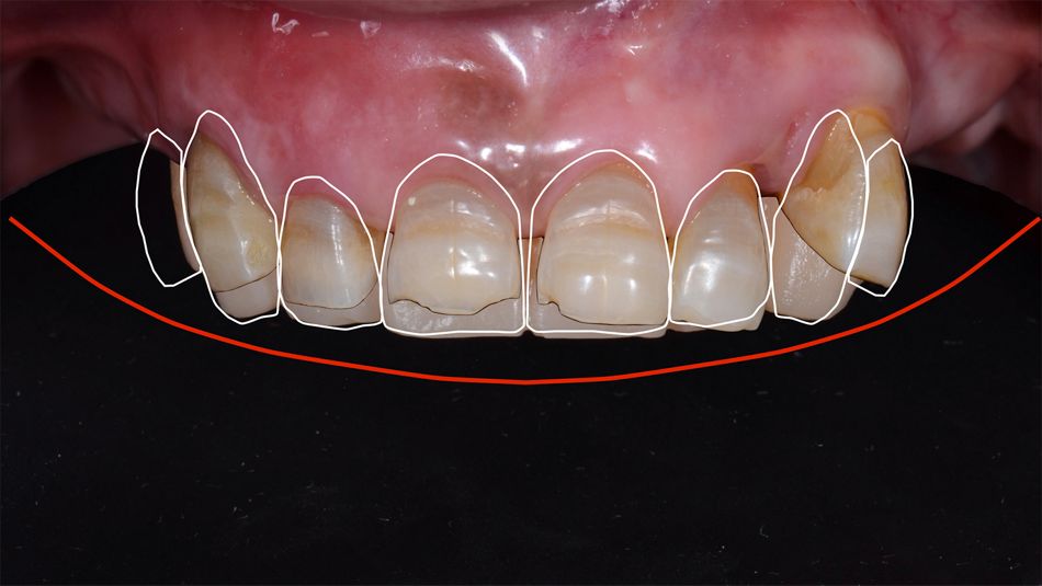
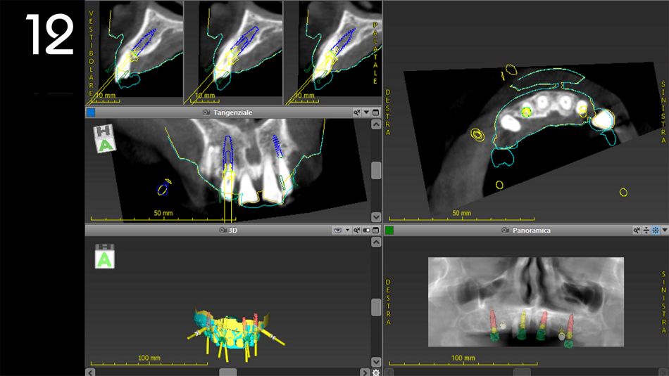
A digital smile design was created and included the initial clinical situation, the proposed shape and position of the teeth, and the lower lip line position (Fig. 12). In addition, the deep bite and the overjet were corrected, and the smile was improved. This prosthetic plan was discussed with the patient and then approved (Figs. 13-15).
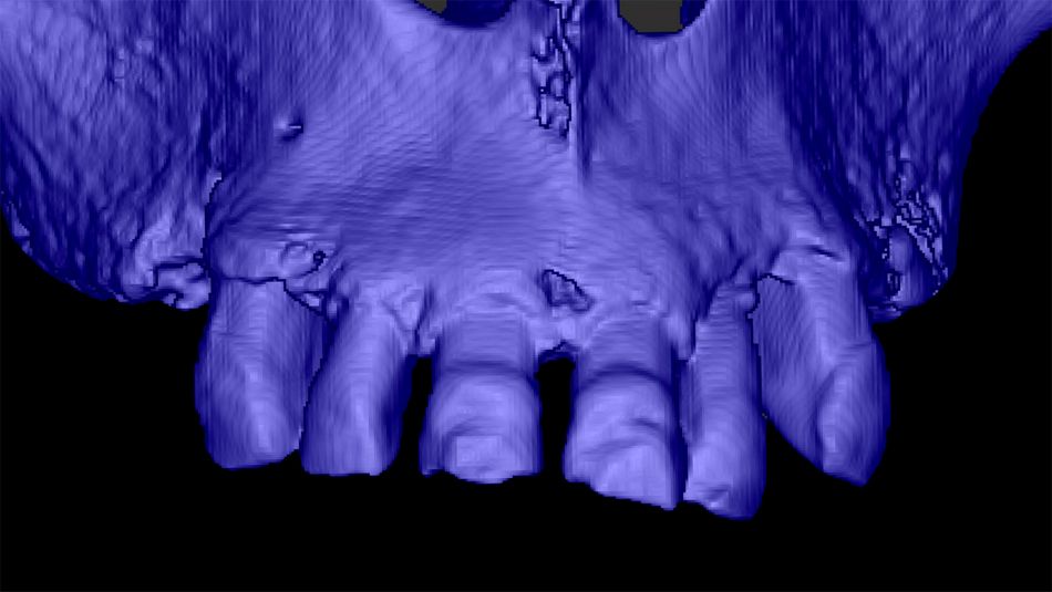
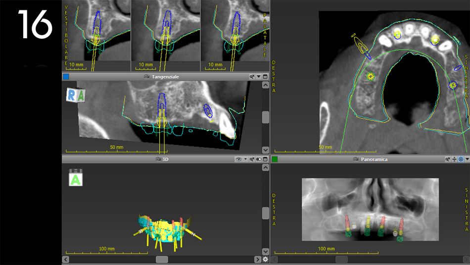
The cast model was scanned and an STL file was generated. Moreover, a CBCT exam was requested for the radiographic assessment. This data, including the DICOM files, were imported in coDiagnostiX® software for the analysis and treatment planning (Fig. 16).
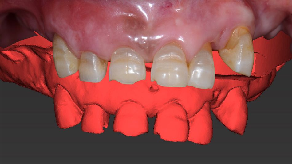
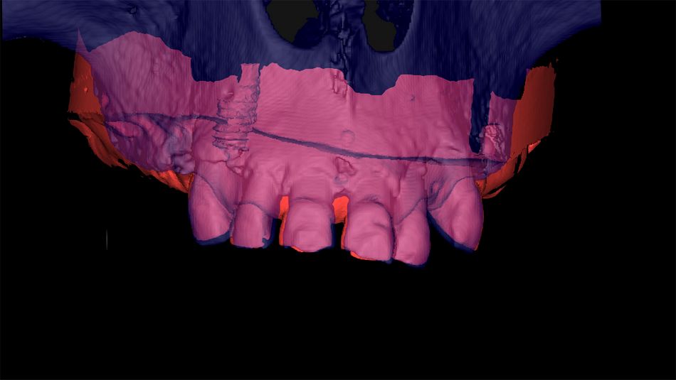
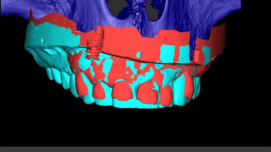
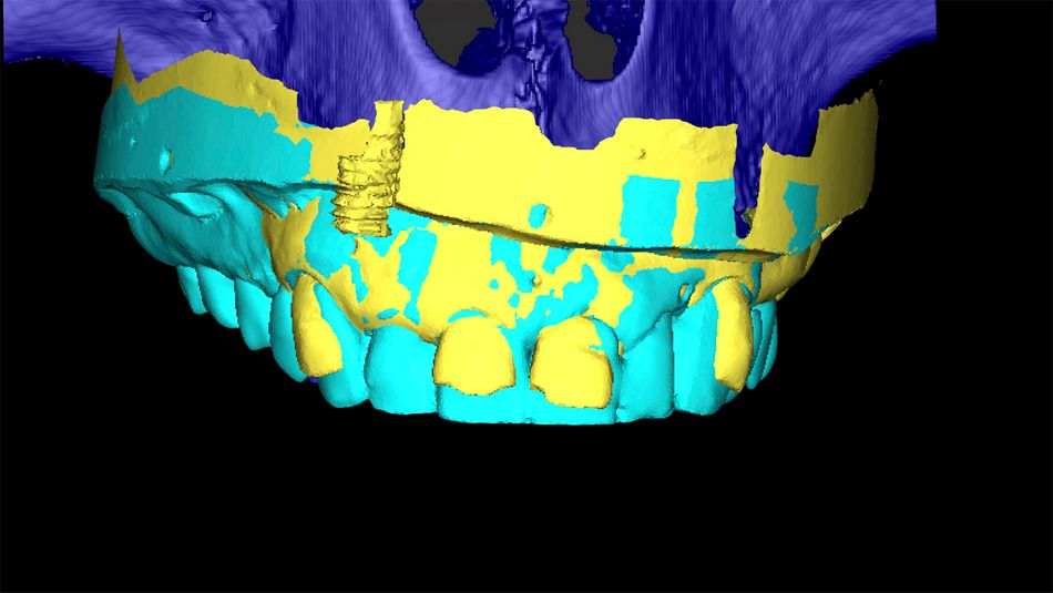
The prosthetic plan is just as important as the surgical plan. For this, the STL file of the initial situation was compared with the DICOM files (Fig. 17,18). Furthermore, the STL file related to the proposed prosthetic plan was aligned with the rest of the files (Fig. 19). Finally, the last pairing was performed by superimposing the STL file with the virtual extraction of the lateral incisors, which were potential sites for implant placement (Fig. 20).
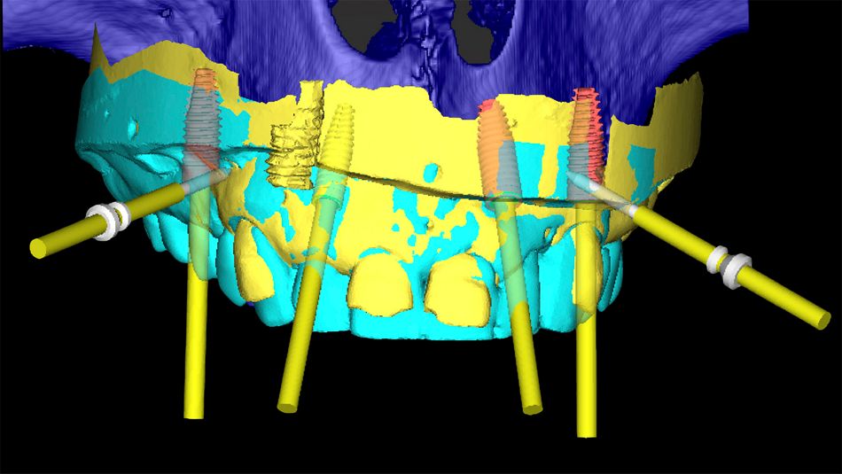
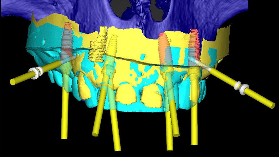
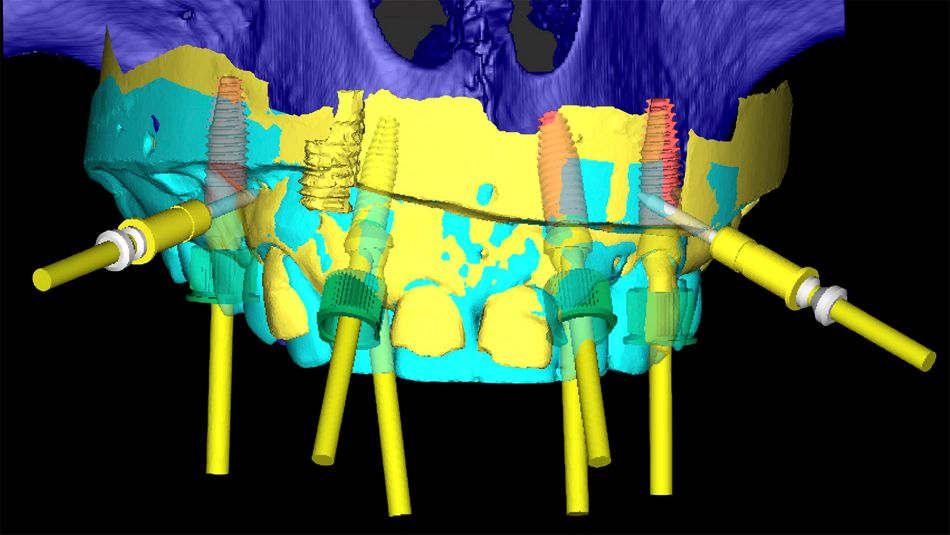
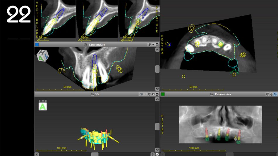
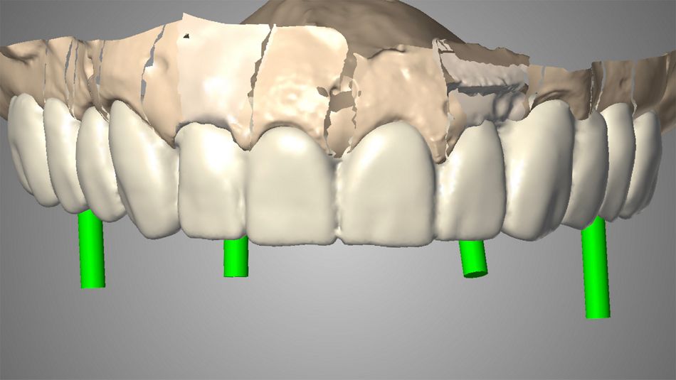
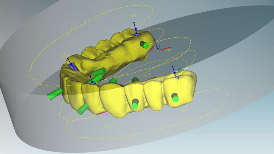
After this preliminary phase, all the data related to the initial situation, bone availability, gingival profiles, and prosthetic strategy were ready to start the planning of implant positioning. On this basis, four Straumann® Bone Level Tapered implants (22: diameter 4.1 length 12; 15: diameter 4.1 length 12; 25: diameter 4.1 length 12; 12: diameter 3.3 length 12) were strategically distributed among the maxilla and fixation pins were considered for the stabilization of the surgical guide (Fig. 21). Furthermore, suitable screw-retained abutments (XXXX) were chosen and the corresponding sleeves for the implant and pin placement were selected (Figs. 22,23).

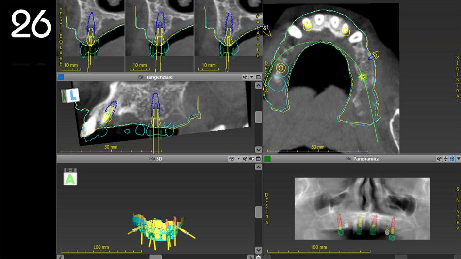
The implants at sites #16, #12, #22, and #26 were planned according to bone volume, soft tissue position, and prosthetic strategy (Figs. 24-27).

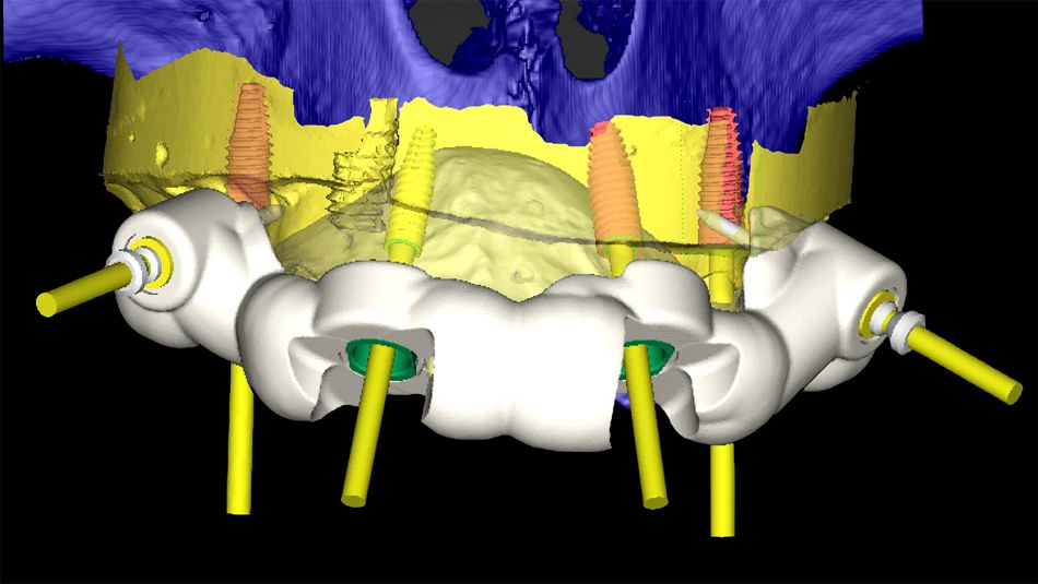
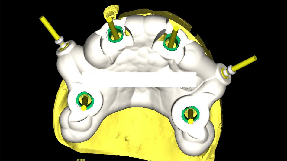
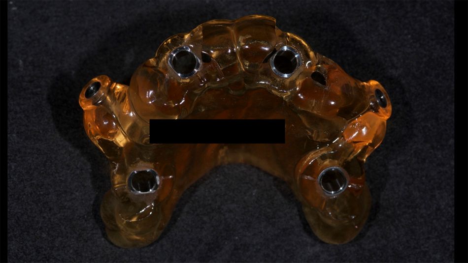
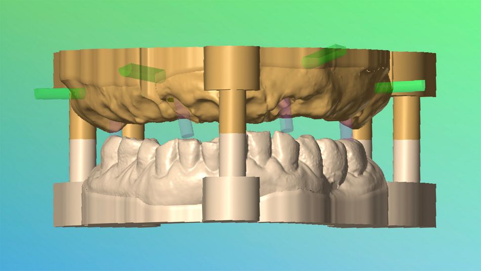
The surgical guide for implant placement was designed and the corresponding STL file was sent to the dental laboratory for printing (Figs. 28-30).

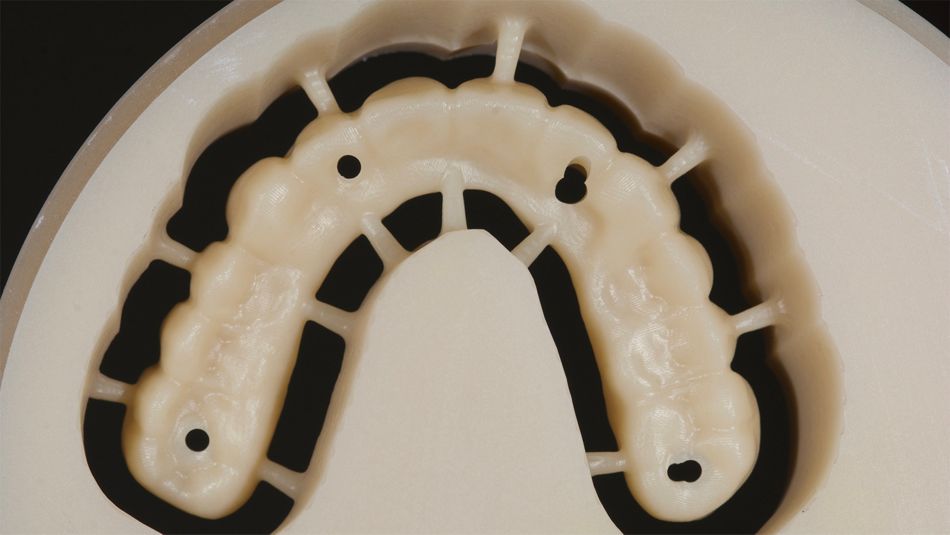
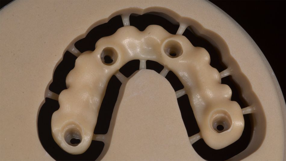
The next step was to prepare the temporary bridge for immediate loading. This is the preliminary phase prior to milling, and involves replicating the shape of the dental set-up in a resin disk. (Figs. 31-33).

Once obtained, the temporary bridge was seated and adapted on the master model with implant analogs and the corresponding SRA abutments (Fig. 34). In the final step, occlusion was carefully adjusted (Fig. 35).

Surgical procedure

In the first phase, the hopeless teeth located on the planned implant sites were strategically extracted, and the soft tissue removed with a punch for a flapless approach. The remaining teeth were used to stabilize the guide with the aid of surgical pins as anchorage.

Following the optimal stabilization of the guide, the implants were placed using the dedicated surgical kit, and the screw-retained abutments (SRA) were placed on the implants.

The remaining teeth on the arch were then extracted and temporary abutments were placed on the SRAs. The temporary bridge was screwed onto the copings and bonded with flowable resin.

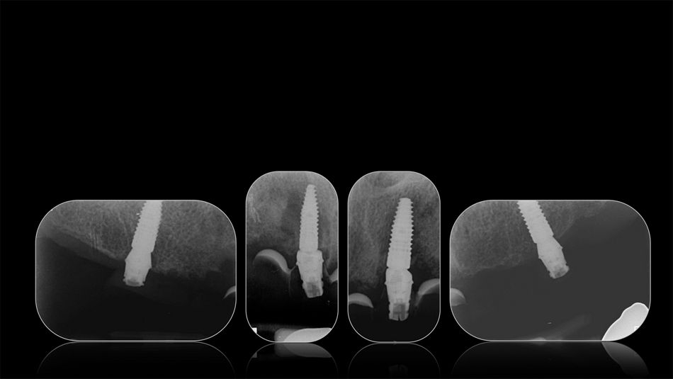
Once the occlusion was adjusted, the chimneys were filled with Teflon and covered with temporary composite. The patient was very satisfied with the functional and esthetic outcome (Fig. 36). X-ray images taken at the one-month (Fig. 37) and at the three-month follow-up visit showed a favorable outcome and the patient reported no mechanical or biological.

Prosthetic procedure

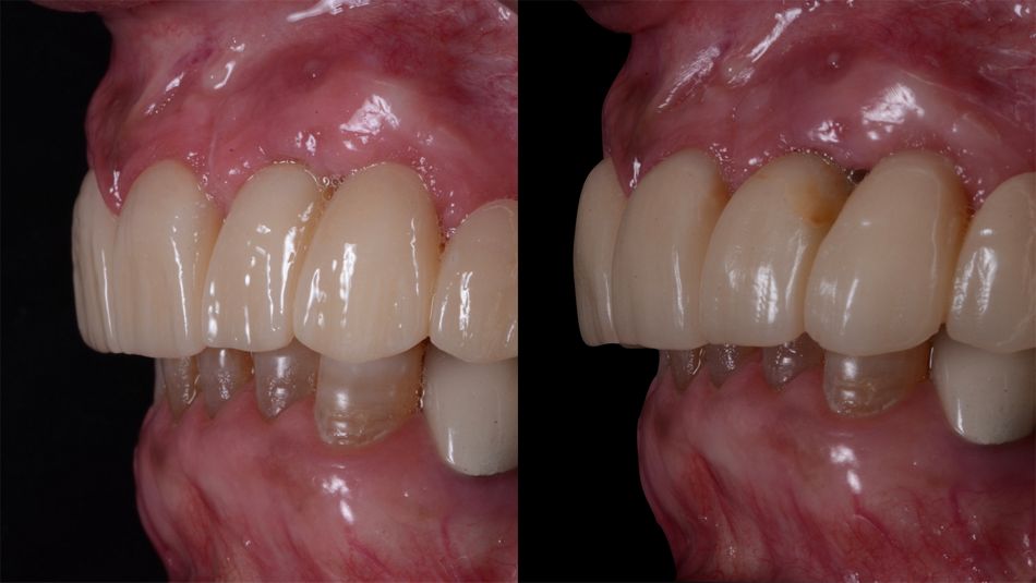
At the 3-month follow-up visit, the patient reported no mechanical or biological complications. Moreover, the clinical examination showed uneventful healing with a complete soft tissue maturation (Fig. 38-41).

At this point, the treatment already met the patient’s expectations providing the desired esthetic and functional clinical outcomes (Fig. 39-40).

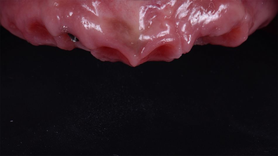
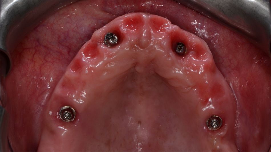
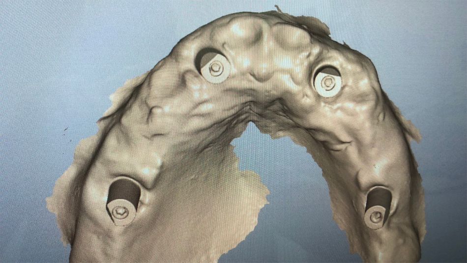
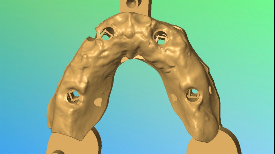
To prepare the final restoration, the temporary bridge was removed, and the soft tissues were evaluated (Fig. 42). An optical impression with Straumann® Virtuo Vivo™ intraoral scanner was taken, and a digital model was obtained and guided into occlusion with the lower jaw (Figs. 43-45).

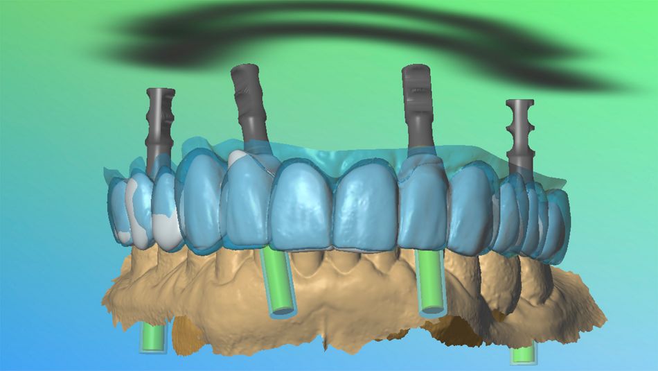
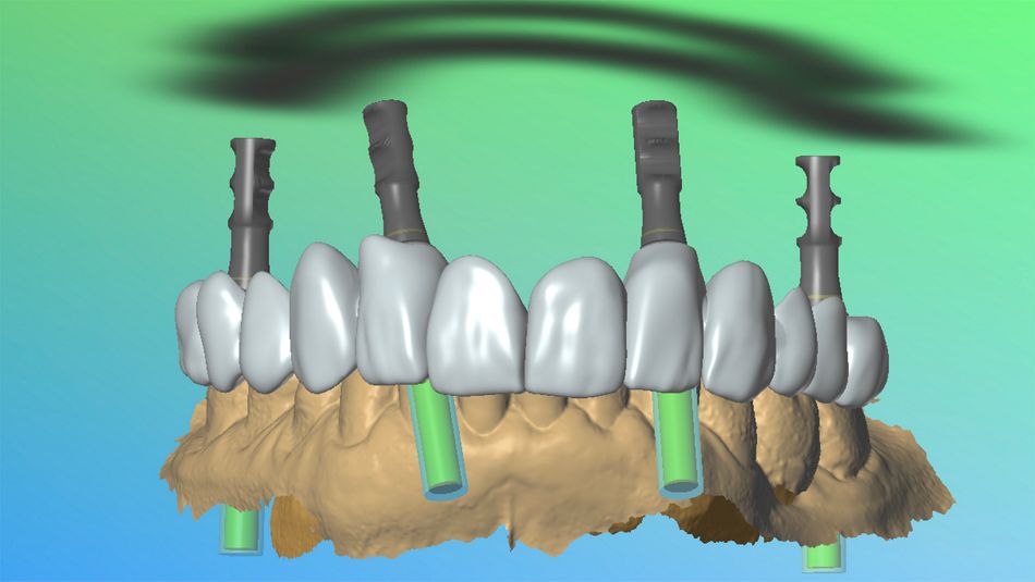
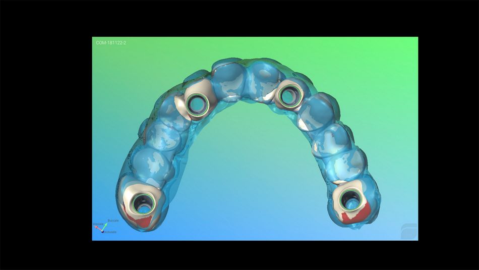
Then, the initial dental set-up (represented in blue in Fig. 46) was matched with the digital model, and minor adjustments were made (shown in white in Fig. 46) for the final dental display (Fig. 47).

Before milling the zirconia framework, it is important to ensure optimal dental optimal dental alignment. Therefore, a “test drive” was performed through a rough milled resin bridge with the exact shape of the designed restoration that was then checked in the patient’s mouth (Figs. 48-50).

The overall esthetic outcome, smile line, emergence profiles of the teeth, lip support, and occlusion were carefully verified.

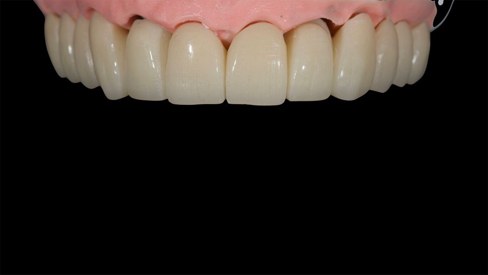
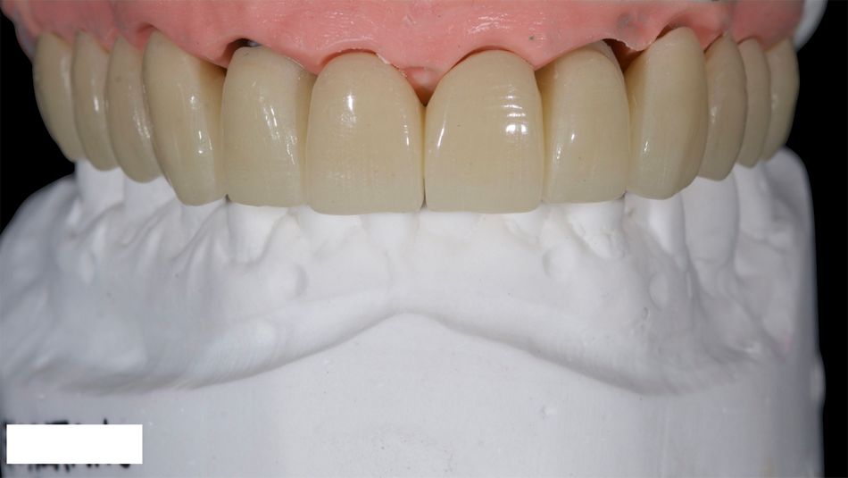
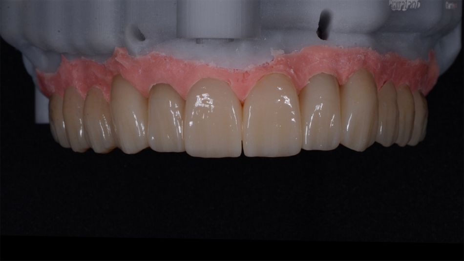
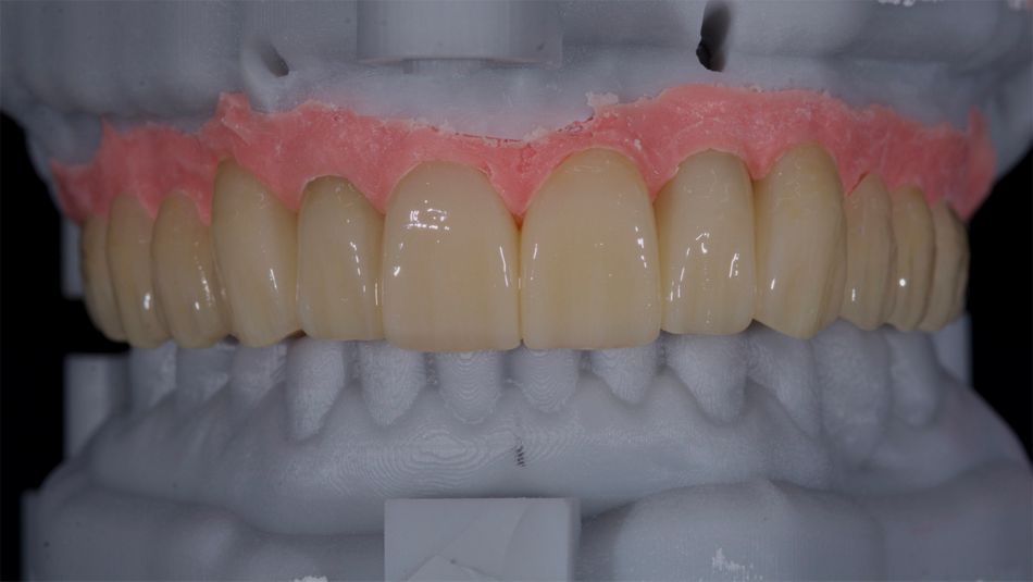
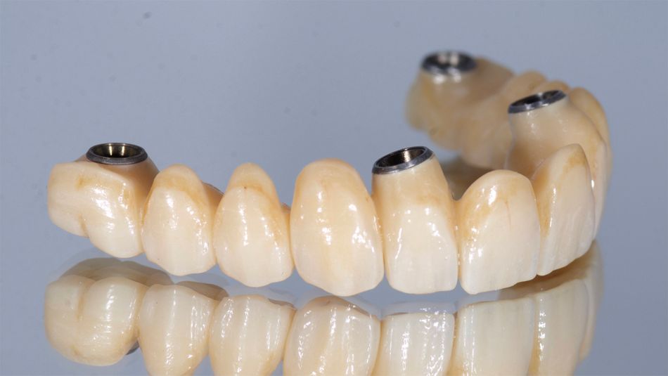
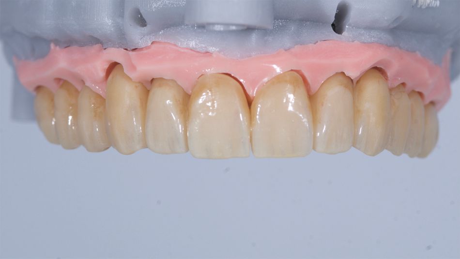
The zirconia bridge was then created accordingly (Figs. 51-55). For this, a full-contour zirconia bridge was milled onto which Variobase® copings were cemented (Fig. 56). The model was slightly adjusted and refined before delivery (Fig. 57).

Responsibility for the outstanding restorations is by dental technician Alessandro Giacometti, who has supported me in this case, and provided a state of the art bridges during the treatment.

Treatment outcomes

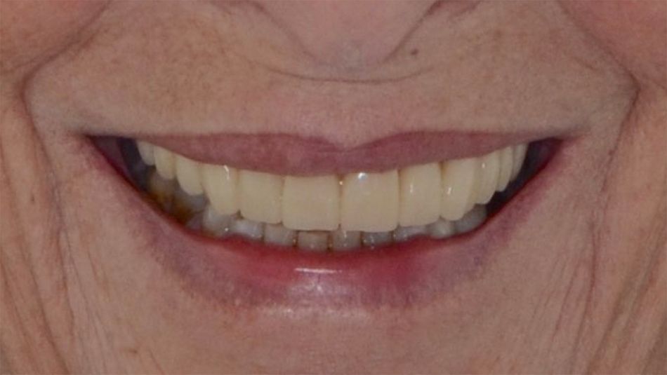
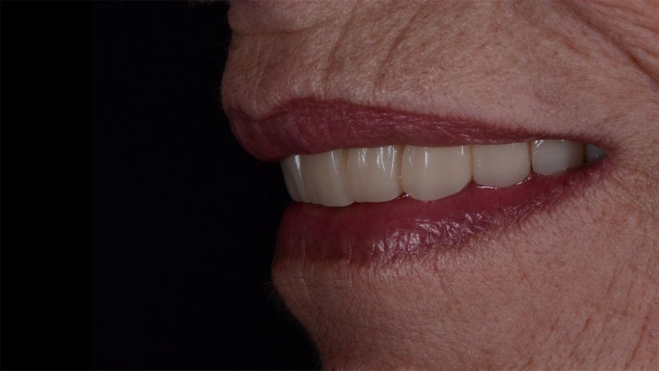
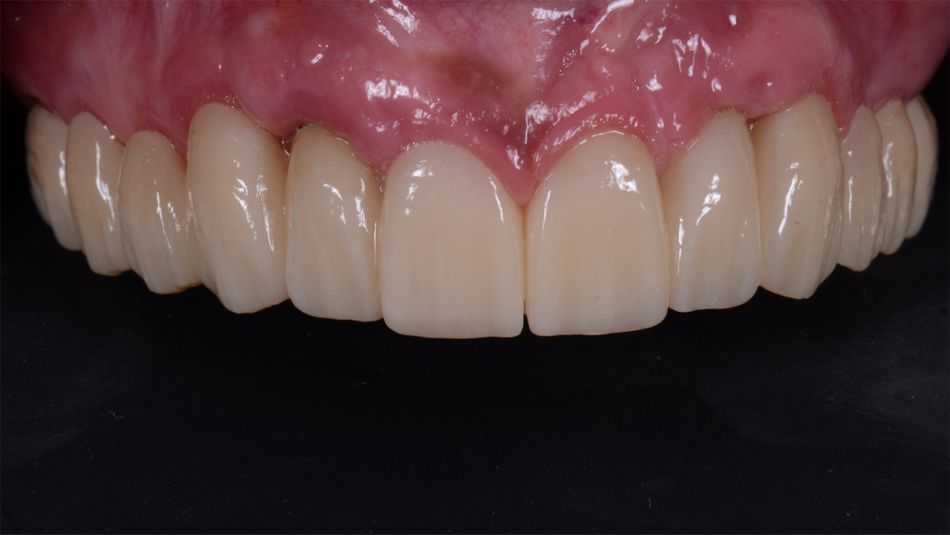
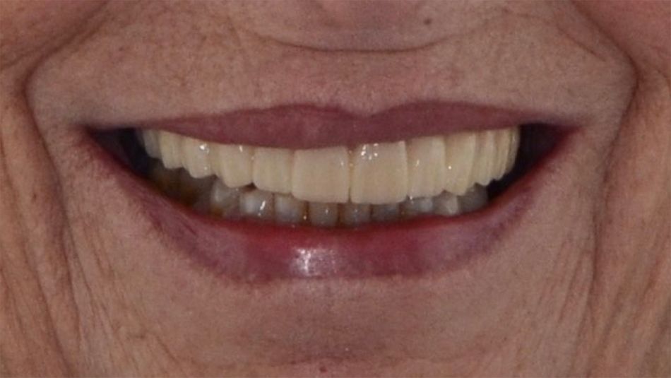
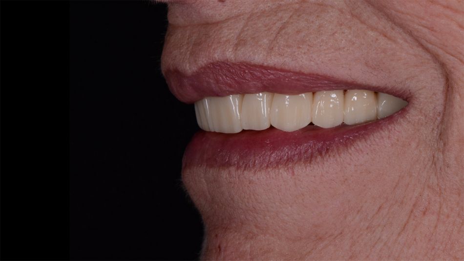
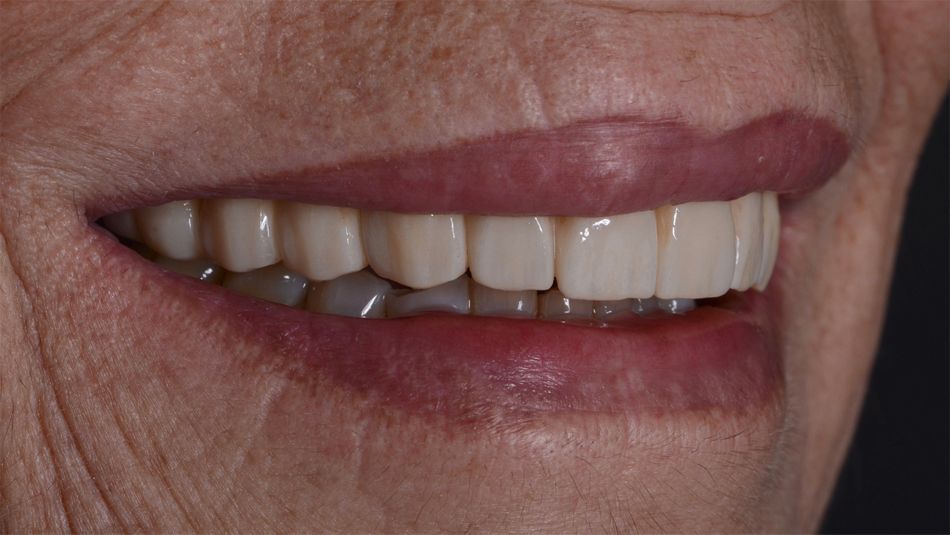
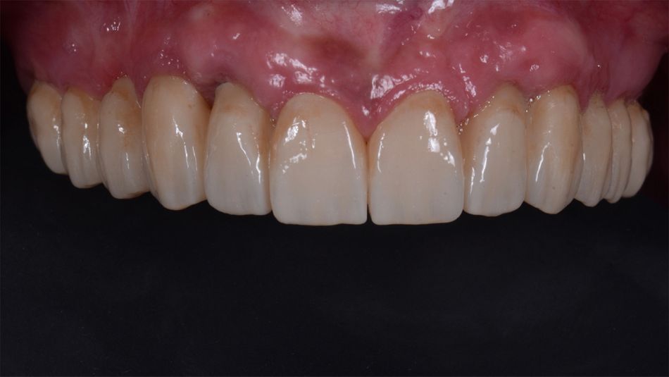
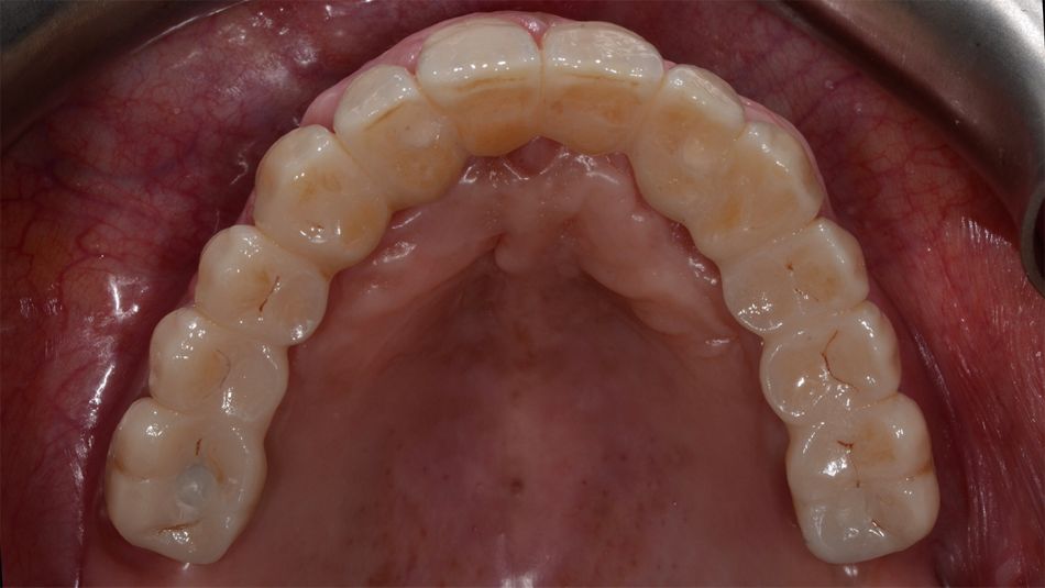
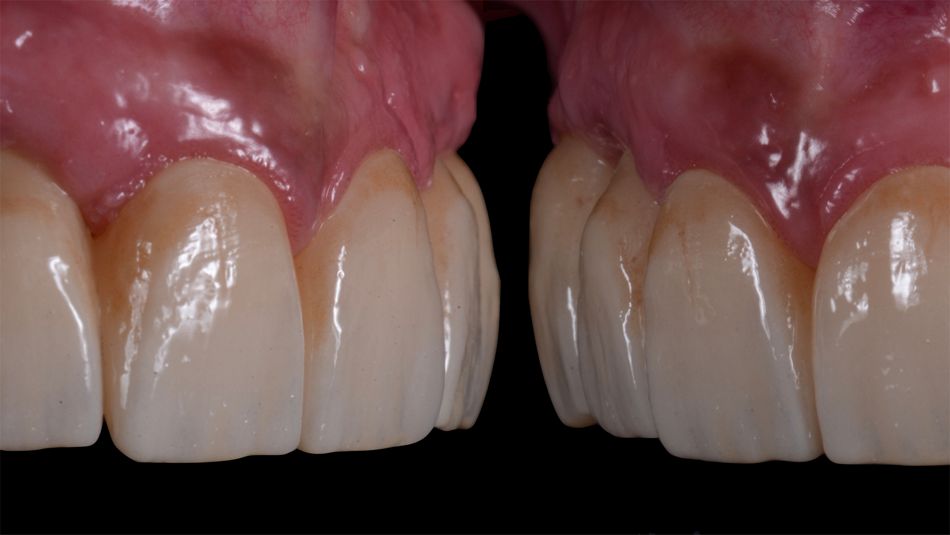
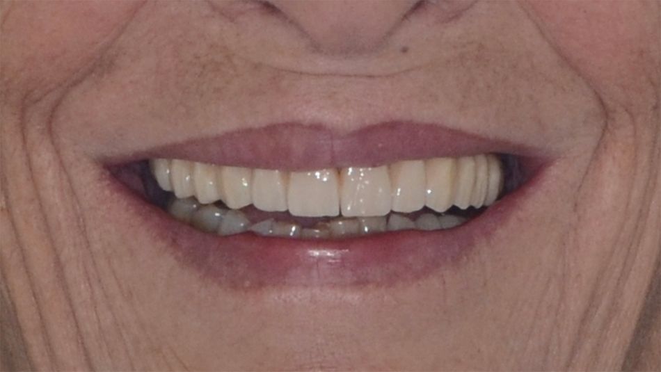
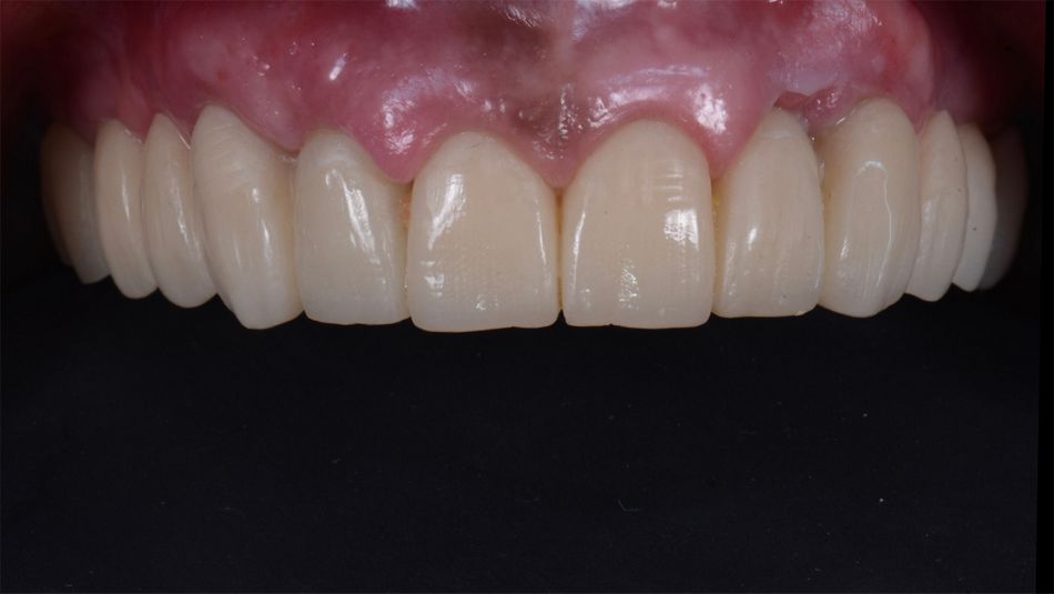
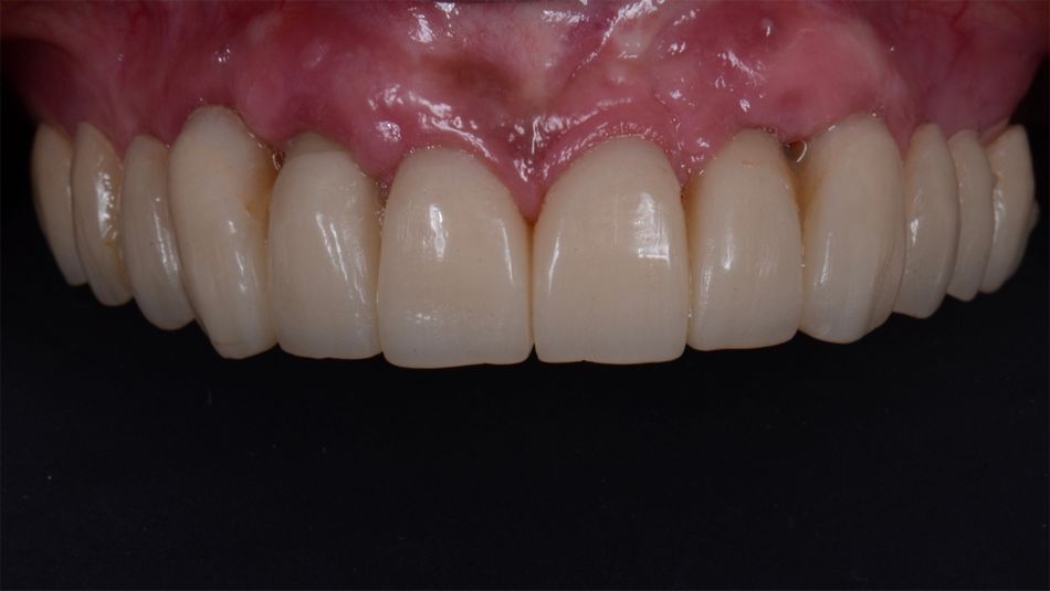
Finally, the bridge was screwed in the patient’s mouth. The treatment outcome fulfilled the patient’s esthetic expectations in terms of natural appearance, harmony, and beauty. Furthermore, she reported an improvement in her quality of life due to the recovery of her masticatory function and self-esteem (Figs 58-62).