#Full-Arch 13. Dec 2021

5 maintenance essentials for the long-term successful fixed full-arch restorations

Dr Andrew Legg, UK

The replacement of a terminal dentition with an implant-supported full arch restorations is a well-established and increasingly endorsed treatment modality [1–3]. A broad range of individual configurations with varying number of implants, placement angles, placement and loading regimens and fixture positioning have been documented to successfully support such rehabilitations [4–9]. Recent research indicates that good clinical long-term success rates of full arch restorations may even be achieved in periodontally compromised patients albeit these patients have been associated with a higher risk for peri-implantitis [10–12].

Dental implants are constantly exposed to the microbe-laden environment of the oral cavity which leads to a rapid formation and growth of biofilms on the prosthetic and connecting elements of the restoration [13]. Supported by appropriate oral hygiene measures these biofilms are held in a stable balance (homeostasis) by the patient’s immune system with the result of clinical peri-implant health.

Peri-implant mucositis and peri-implantitis represent undesired biological complications that may ultimately lead to implant loss and treatment failures14. Restorative designs, treatment regimens and maintenance protocols have to ensure that conditions can be achieved and maintained that successfully prevent such complications. Here we present 5 simple, but fundamental aspects that should be considered in your clinical practice to ensure the long-term success of full arch restorations.

1. Start at the beginning - Presurgical hygiene

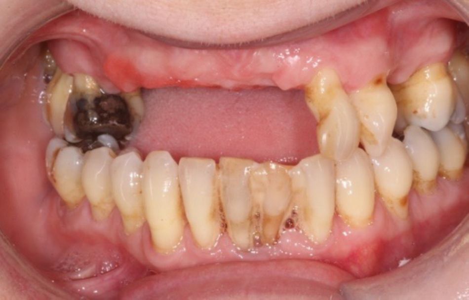
Patients considered for full-arch restorations often present with a failing dentition affected by severely progressed caries and periodontitis. These clinical findings are often associated to a history of poor oral hygiene skills and habits. Implant-placement in such patients seems to be counter-intuitive as any reoccurrence of such pathogen associated conditions may represent a risk-factor to the future implant restoration. Therefore, the reestablishment of an adequate oral health status is as important as the instillation of the fundamentals and habits of good oral hygiene into patients prior to commencing any advanced implant treatment.

Along and following causative treatment of any pathogenic conditions patients may be prepared for implant therapy by thorough instructions for oral hygiene. Reevaluation visit may e.g. not only be used to assess the patient’s response to treatments but also to ensure that adequate levels of plaque control are achieved and maintained by compliance with oral hygiene routines14.

Further treatment planning should comprise a thoughtful analysis of risk factors related to e.g. the recurrence of periodontal disease or the onset of any future peri-implant disease. As a general guideline, any precondition that may interfere or alter the innate or adaptive immune responses to a bacterial challenge, like. e.g. smoking may put the oral health status and any future implant restoration at risk15, 16.

The implementation of an early preoperative hygiene therapy regimen represents an integral part to our treatment approach at Campbell clinic when providing full arch restorations. We routinely provide patient individualized oral hygiene instruction and constantly evaluate patient compliance with oral hygiene principles to improve post-operative outcomes and the results of our maintenance regimes17–19.

2. Plan carefully- consider hygiene and maintenance aspects of implant and prosthetic restorative designs

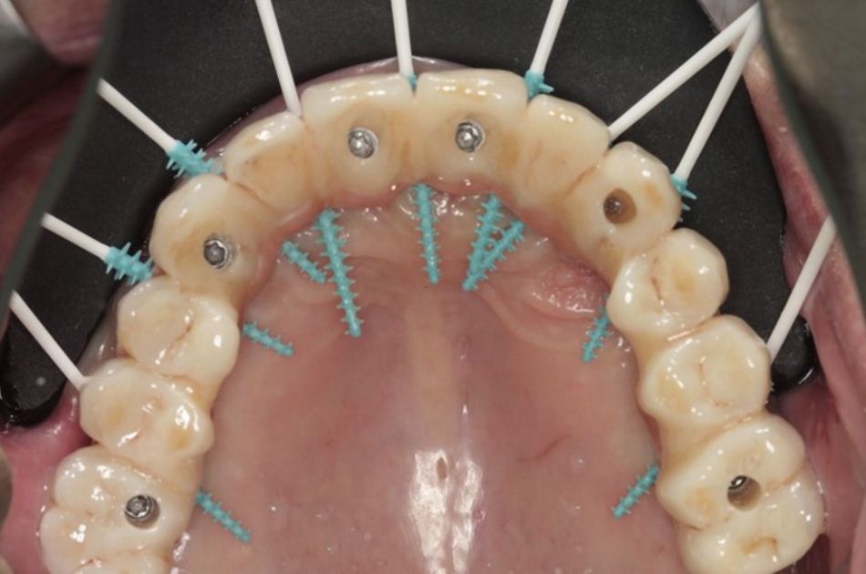
The prosthetic design is critical to any full arch reconstruction. Besides mechanical and esthetics aspects the ability to successfully allow for adequate plaque control should be considered already at the planning stage of the implant and prosthetic restorations. Full arch restorations should be planned with the aim to limit the accumulation and allow a facile removal of pathogenic bacteria. Furthermore, patient specific risk factors for peri-implantitis as well as individual skills and abilities to clean may vary from patient to patient20, 21. Such patient-specific factors should be taken into consideration when planning the hygienic aspects and requirements of the individual full arch restorative design.

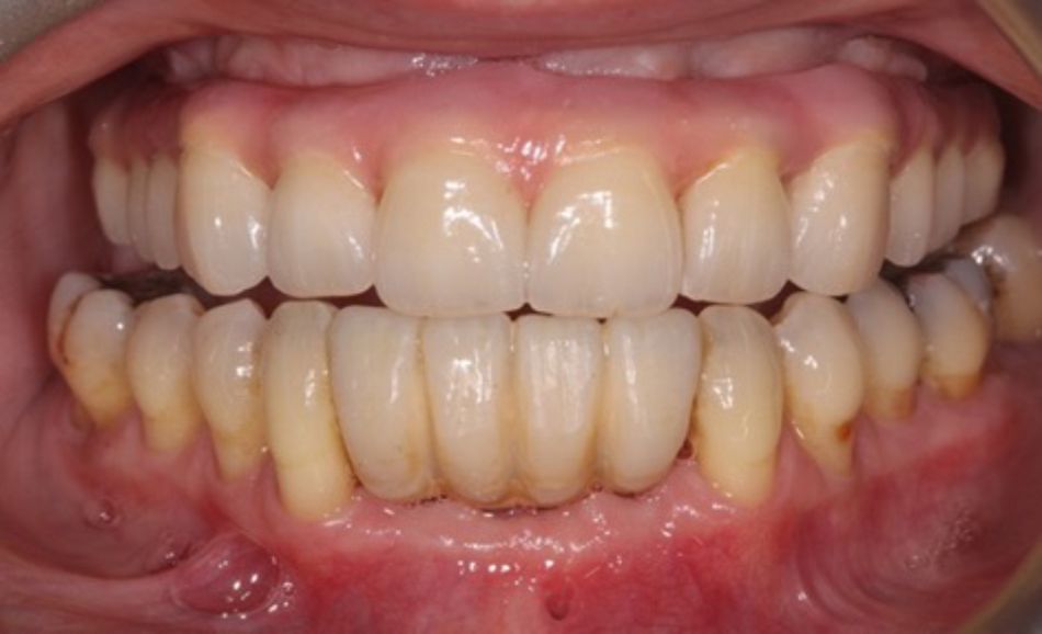
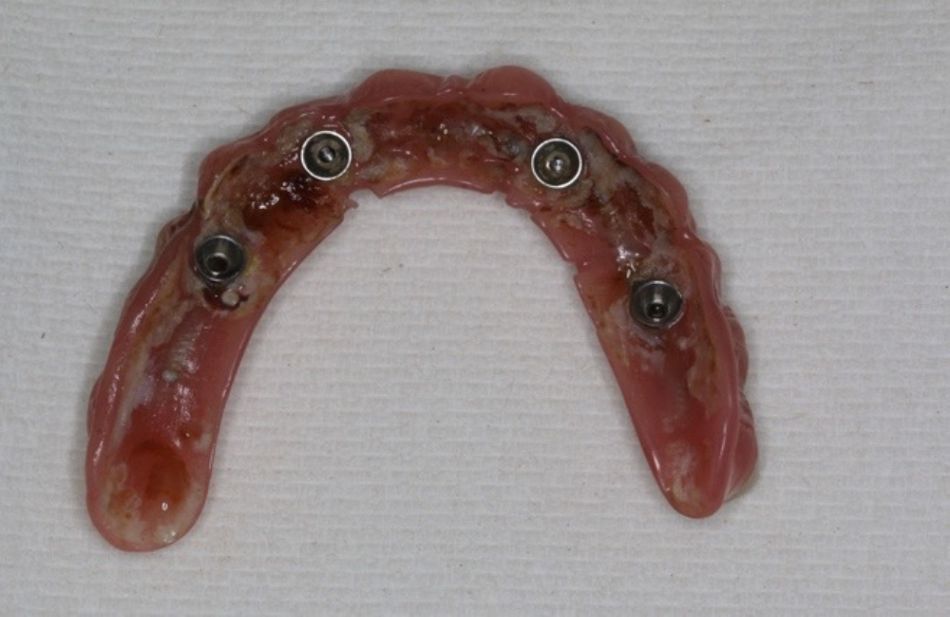
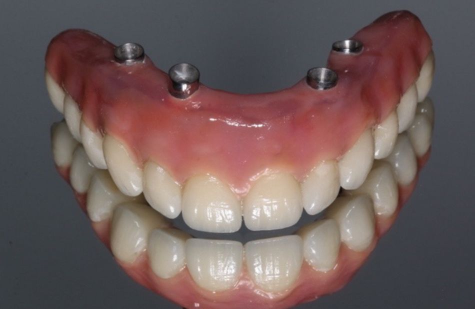
Depending on the available restorative space and esthetic requirements the prosthetic cervical and specifically pontic and connecting designs of the prosthetic framework can be individually and even locally adapted to meet the requirements for cleaning and maintenance22. Access for cleaning needs also to be considered when planning the spacing between implants. Inter-implant pontic sections should remain accessible for cleaning. Implant emergence and pontic profiles should be designed with hygenic, i.e. preferably ovate contours (Fig. 4). Ridge lap pontic designs should whenever possible be avoided for the design of fixed full arch restorations as they are associated with an elevated risk for food entrapment and plaque accumulation while impeding the ability to clean (Fig.3)23. Lastly whenever possible screw retained restorations should be used, which facilitates the management of any mechanical and biological complications after prosthetic delivery.

3. Advice properly - Post-Surgical Instructions

Post-surgical instructions for adequate homecare are essential to support the healing of implant sites in full arch restorations, specifically when applying immediate loading regimens. These instructions should be related to both oral care, food intake and inform the patients on the specific conditions that may require immediate assistance.

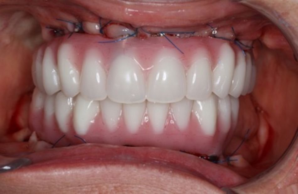
Specifically, in the case of immediately loaded full arch restorations more careful instructions may be need. Such instructions should restrict or prevent mechanical cleaning routines that may be adequate for routine cleaning, like e.g. the use of air or water jet devices. Patients may be instructed to apply specific brushing techniques, e.g. in a vertical, apical direction to support the soft tissue adaptation to the prosthetic framework, however without compromising the provisional soft tissue fixation around the implants (Fig. 5). Chlorhexidine mouthwashes may be used to support plaque control during this period. Also depending on the specific individual indication patients may be advised to refer to soft diet only for the first weeks after surgery.

In the case of zygomatic implants, all pressured cleaning devices would be avoided due to the tenuous nature of the soft tissue connection around the implants. These patients should be advised to carry out manual and very careful cleaning only.

Patients further may need to be sensitized that a series of conditions like e.g. excessive bleeding, severe pain etc. may occur that require immediate assistance by the treating clinician.


Want to stay up to date?

youTooth.com is THE PLACE TO BE IN DENTISTRY – subscribe now and receive our monthly newsletter on top hot topics from the world of modern dentistry.


4. Remain flexible and adapt your approach to cleaning

Adequate self-administered and professional oral hygiene routines are crucial to ensure proper plaque control and to limit the accumulation of pathogenic bacteria causing peri-implant inflammation. Such routines may comprise brushing at least twice daily, use of floss, interdental cleaners, and water irrigators and/or a combination of these measures.

It is essential that clinicians, both dentists and hygienists, are flexible in their approach to full arch maintenance, which may require to establish a patient-specific program of home and professional care. Besides considering patient-specific conditions, like e.g. age, frailty and comorbidities when designing the restoration clinicians and patients may need to individualize cleaning routines to the requirements and capabilities of the corresponding patient. Some patients will e.g. be able to use bridge floss, while elderly or frail patients may only be able to use interdental brushes or powered cleaning devices. Following strict dogmatic and rigid approaches to cleaning may unsettle, discourage and ultimately disenfranchise patients with less dexterity. Hence, clinicians need to individualize instructions and assist patients in applying appropriate routines to ensure successful maintenance of their full arch restorations.

Lastly the setup and implementation of patient-specific and individualized approaches to oral care may be facilitated by a seamless communication between clinicians, i.e. dentists and hygienists that should include both technical details related to the specific type of restoration as well as to the details of in office and home care protocols.

Courtesy Dr Alessandro Perucci, Switzerland

5. Make it a routine- Set out a strict hygiene schedule

The maintenance of peri-implant soft tissue health is of utmost importance to ensure long-term success of full arch rehabilitations. Well-designed supportive implant therapy programs with periodic in-office recall visits represent an integral part of full arch implant therapy18. The primary aim of these recalls is to prevent the onset of biological complications, i.e. peri-implant mucositis and peri-implantitis. This is achieved by thorough examination of implant health related parameters and mechanical fit, by applying professional cleaning and by reconfirming and reinforcing home care routines24. Professional cleaning routines represent important elements to ensure the complete removal of any residual supra- and subgingival microbial deposits. The use of instruments, tools or agents that may scratch and roughen the implant or abutments surfaces should be prevented as this may increase the tendency for plaque accumulation as potential source of inflammation25. A recently introduced chitosan brush (Labrida BioCleanTM) represents an attractive candidate that enables the soft and gentle cleaning of the implant surface without showing the typical risks for pro-inflammatory plastic remnants associate to e.g. plastic curettes26, 27.

A regular assessment of the diagnostic parameters can help to identify any biological complications early. Diagnostic parameters may comprise visual analysis for any signs of redness, swelling as well as changes in probing depth, bleeding on probing and suppuration. Depending on the individual design of the prosthetic restoration probing under fixed full arch restorations might be difficult. Nevertheless, the aim should be to identify at least one surface per implant where probing can be performed28.

We routinely start with an in-office implant maintenance interval of 3 months which may be individually adapted and can be shortened or even prolonged based on the patient’s oral hygiene status, compliance with regular home care routines and which can be tailored to the patient’s risk profile. The latter can be usually assessed by evaluating e.g. any history of smoking, periodontitis, systemic conditions as well as the patient’s capabilities and dexterity to clean. Such profiles may change in time and it is advised to regularly update corresponding evaluations and to confirm or readapt intervals and regimens over the course of time. It is important to generate awareness for and to openly communicate the importance of supportive implant therapy programs and the associated time and financial efforts associated to it at the very beginning of implant therapy. Patients may need to be prepared for and willing to commit to supportive therapy for the lifetime of the restoration. Additionally, the standard of care comprises to take radiographs every 1 to 2 years and to compare them to baseline radiographs in order to exclude any changes of the peri-implant bone and to reconfirm firm and adequate passive fit of the prosthetic superstructure to the implant base and connecting parts.

Finally, in our practice, we use to remove the prosthesis every 2 years or otherwise in case of any mechanical or biological complication that may not be solved without removal. This allows for a thorough assessment and direct cleaning of the abutments and implants. Abutment and implant screws are checked and may be exchanged or retightened only if necessary. The prosthesis can be cleaned in antiseptic solution in an ultrasonic bath to remove the biofilm, the screws should be exchanged. It is important to note that this is the protocol in our clinic and other opinions (such as the AAP) suggest leaving the bridge in place unless there is a complication. It is up to the individual clinician to make their own judgement and it is clear that further research is required to form a solid consensus.

Conclusions

Full arch restorations represent a successful treatment modality for the rehabilitation of edentulous patients. Maintenance and implant supportive therapy represents an integral part of implant therapy that is indispensable to ensure the long-term success of such restorations. Aspects for maintenance need to be individually assessed and patient-specifically adapted and considered during the design of the restoration, in the instructions for home care routines and in in-office recall regimens.

References:

  1. Rohlin M, Dr O, Nilner K, et al (2012) Treatment of Adult Patients with Edentulous Arches: A Systematic Review. The International Journal of Prosthodontics 25:553–567
  2. Van Der Bilt A, Burgers M, Van Kampen FMC, Cune MS (2010) Mandibular implant-supported overdentures and oral function: Overdentures and oral function. Clinical Oral Implants Research 21:1209–1213. https://doi.org/10.1111/j.1600-0501.2010.01915.x
  3. Shigehara S, Ohba S, Nakashima K, et al (2015) Immediate Loading of Dental Implants Inserted in Edentulous Maxillas and Mandibles: 5-Year Results of a Clinical Study. Journal of Oral Implantology 41:701–705. https://doi.org/10.1563/AAID-JOI-D-14-00018
  4. Daudt Polido W, Aghaloo T, Emmett TW, et al (2018) Number of implants placed for complete‐arch fixed prostheses: A systematic review and meta‐analysis. Clin Oral Impl Res 29:154–183. https://doi.org/10.1111/clr.13312
  5. Menini M, Signori A, Tealdo T, et al (2012) Tilted implants in the immediate loading rehabilitation of the maxilla: a systematic review. J Dent Res 91:821–827. https://doi.org/10.1177/0022034512455802
  6. Agliardi E (2010) Immediate rehabilitation of the edentulous jaws with full fixed prostheses supported by four implants: interim results of a single cohort prospective study. 7
  7. Pera P, Menini M, Pesce P, et al (2018) Immediate Versus Delayed Loading of Dental Implants Supporting Fixed Full-Arch Maxillary Prostheses: A 10-year Follow-up Report. Int J Prosthodont 32:27–31. https://doi.org/10.11607/ijp.5804
  8. Papaspyridakos P, Chen C-J, Chuang S-K, Weber H-P (2014) Implant Loading Protocols for Edentulous Patients with Fixed Prostheses: A Systematic Review and Meta-Analysis.
  9. Papaspyridakos P, Mokti M, Chen C-J, et al (2014) Implant and Prosthodontic Survival Rates with Implant Fixed Complete Dental Prostheses in the Edentulous Mandible after at Least 5 Years: A Systematic Review: Implant and Prosthesis Survival Rates in Edentulous Mandible. Clinical Implant Dentistry and Related Research 16:705–717. https://doi.org/10.1111/cid.12036
  10. Alves CC, Correia AR, Neves M (2010) Immediate implants and immediate loading in periodontally compromised patients-a 3-year prospective clinical study. Int J Periodontics Restorative Dent 30:447–455
  11. Li S, Di P, Zhang Y, Lin Y (2017) Immediate implant and rehabilitation based on All-on-4 concept in patients with generalized aggressive periodontitis: A medium-term prospective study: Li et al. Clinical Implant Dentistry and Related Research 19:559–571. https://doi.org/10.1111/cid.12483
  12. Chrcanovic BR, Albrektsson T, Wennerberg A (2014) Periodontally compromised vs. periodontally healthy patients and dental implants: A systematic review and meta-analysis. Journal of Dentistry 42:1509–1527. https://doi.org/10.1016/j.jdent.2014.09.013
  13. Subramani K, Jung RE, Molenberg A, Hammerle CHF (2009) Biofilm on dental implants: a review of the literature. Int J Oral Maxillofac Implants 24:616–626
  14. Armitage GC, Xenoudi P (2016) Post-treatment supportive care for the natural dentition and dental implants. Periodontol 2000 71:164–184. https://doi.org/10.1111/prd.12122
  15. Kinane DF, Demuth DR, Gorr S-U, et al (2007) Human variability in innate immunity. Periodontol 2000 45:14–34. https://doi.org/10.1111/j.1600-0757.2007.00220.x
  16. Chrcanovic BR, Albrektsson T, Wennerberg A (2015) Smoking and dental implants: A systematic review and meta-analysis. Journal of Dentistry 43:487–498. https://doi.org/10.1016/j.jdent.2015.03.003
  17. Corbella S, Del Fabbro M, Taschieri S, et al (2011) Clinical evaluation of an implant maintenance protocol for the prevention of peri-implant diseases in patients treated with immediately loaded full-arch rehabilitations: Implant maintenance protocol for immediate loading. International Journal of Dental Hygiene 9:216–222. https://doi.org/10.1111/j.1601-5037.2010.00489.x
  18. Costa FO, Takenaka-Martinez S, Cota LOM, et al (2012) Peri-implant disease in subjects with and without preventive maintenance: a 5-year follow-up. J Clin Periodontol 39:173–181. https://doi.org/10.1111/j.1600-051X.2011.01819.x
  19. Quirynen M, Abarca M, Van Assche N, et al (2007) Impact of supportive periodontal therapy and implant surface roughness on implant outcome in patients with a history of periodontitis. J Clin Periodontol 34:805–815. https://doi.org/10.1111/j.1600-051X.2007.01106.x
  20. Chrcanovic BR, Albrektsson T, Wennerberg A (2014) Diabetes and Oral Implant Failure: A Systematic Review. J Dent Res 93:859–867. https://doi.org/10.1177/0022034514538820
  21. Romandini M, Lima C, Pedrinaci I, et al (2021) Prevalence and risk/protective indicators of peri‐implant diseases: A university‐representative cross‐sectional study. Clin Oral Impl Res 32:112–122. https://doi.org/10.1111/clr.13684
  22. Bedrossian E, Sullivan RM, Fortin Y, et al (2008) Fixed-Prosthetic Implant Restoration of the Edentulous Maxilla: A Systematic Pretreatment Evaluation Method. Journal of Oral and Maxillofacial Surgery 66:112–122. https://doi.org/10.1016/j.joms.2007.06.687
  23. Steven C-L Use of a Modified Ovate Pontic in Areas of Ridge Defects: A Report of Two Cases. 11
  24. Monje A, Aranda L, Diaz KT, et al (2016) Impact of Maintenance Therapy for the Prevention of Peri-implant Diseases: A Systematic Review and Meta-analysis. J Dent Res 95:372–379. https://doi.org/10.1177/0022034515622432
  25. Mengel R, Buns CE, Mengel C, Flores-de-Jacoby L (1998) An in vitro study of the treatment of implant surfaces with different instruments. Int J Oral Maxillofac Implants 13:91–96
  26. Mann M, Parmar D, Walmsley AD, Lea SC (2012) Effect of plastic-covered ultrasonic scalers on titanium implant surfaces: Modified ultrasonic scalers and implant surfaces. Clinical Oral Implants Research 23:76–82. https://doi.org/10.1111/j.1600-0501.2011.02186.x
  27. Wohlfahrt JC, Evensen BJ, Zeza B, et al (2017) A novel non-surgical method for mild peri-implantitis- a multicenter consecutive case series. Int J Implant Dent 3:38. https://doi.org/10.1186/s40729-017-0098-y
  28. Lindhe J, Meyle J, on behalf of Group D of the European Workshop on Periodontology (2008) Peri-implant diseases: Consensus Report of the Sixth European Workshop on Periodontology. Journal of Clinical Periodontology 35:282–285. https://doi.org/10.1111/j.1600-051X.2008.01283.x