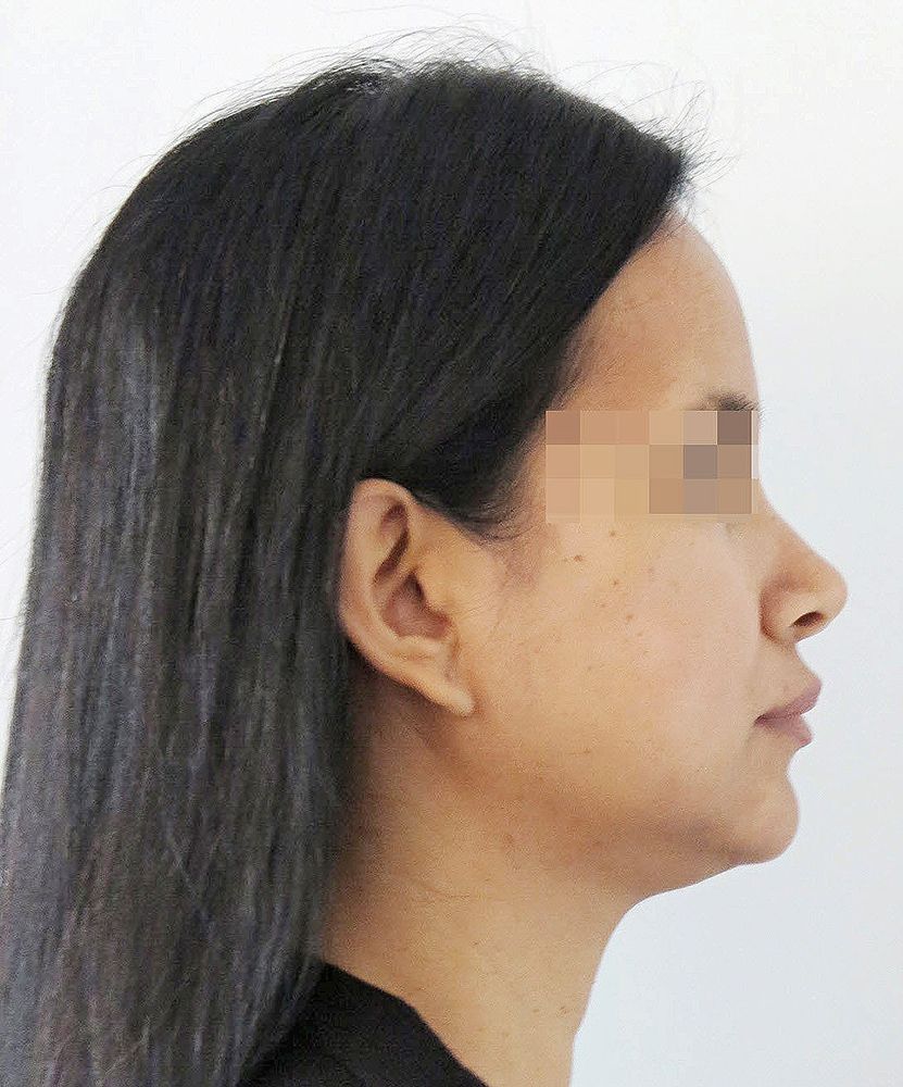
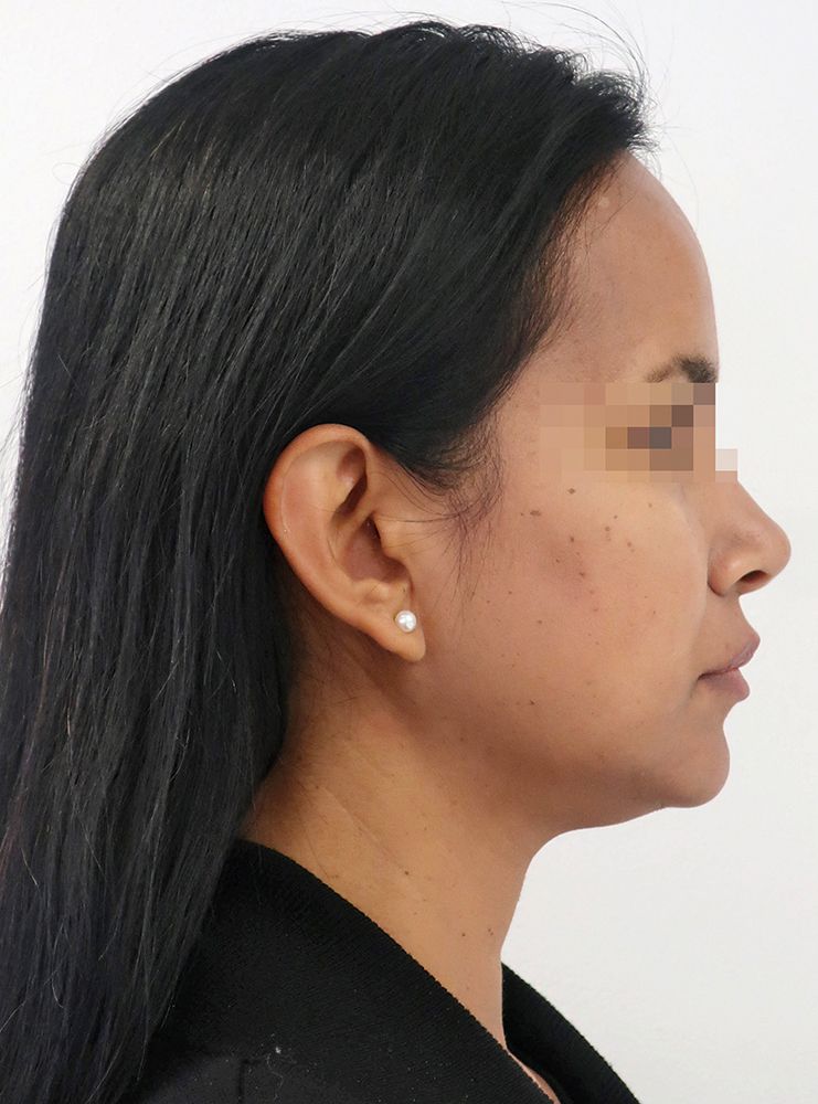
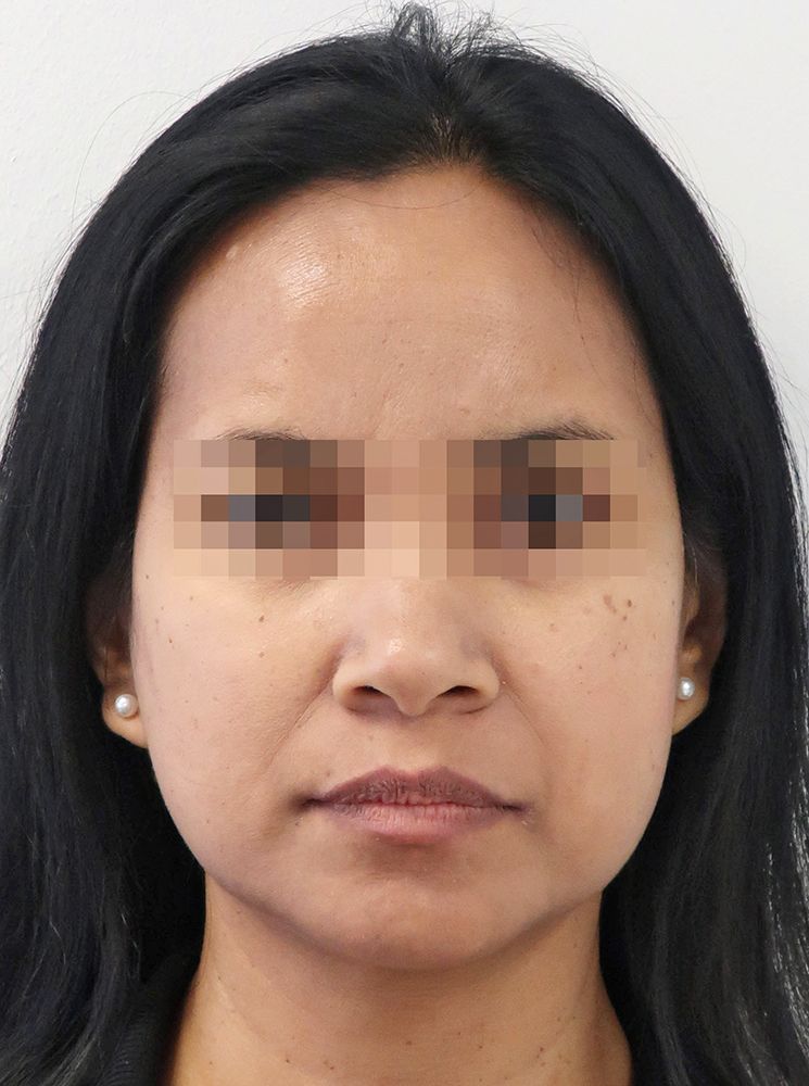
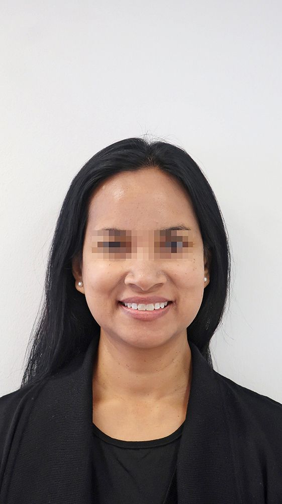
- Figs. 1a: Initial extraoral patient photos, front and profile
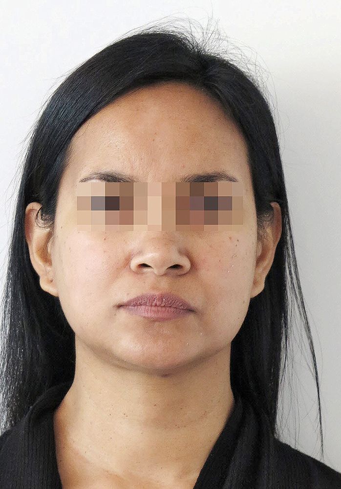
- Figs. 1b: Initial extraoral patient photos, front and profile
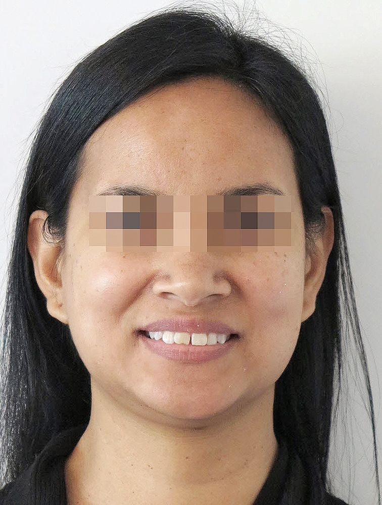
- Figs. 1c: Initial extraoral patient photos, front and profile
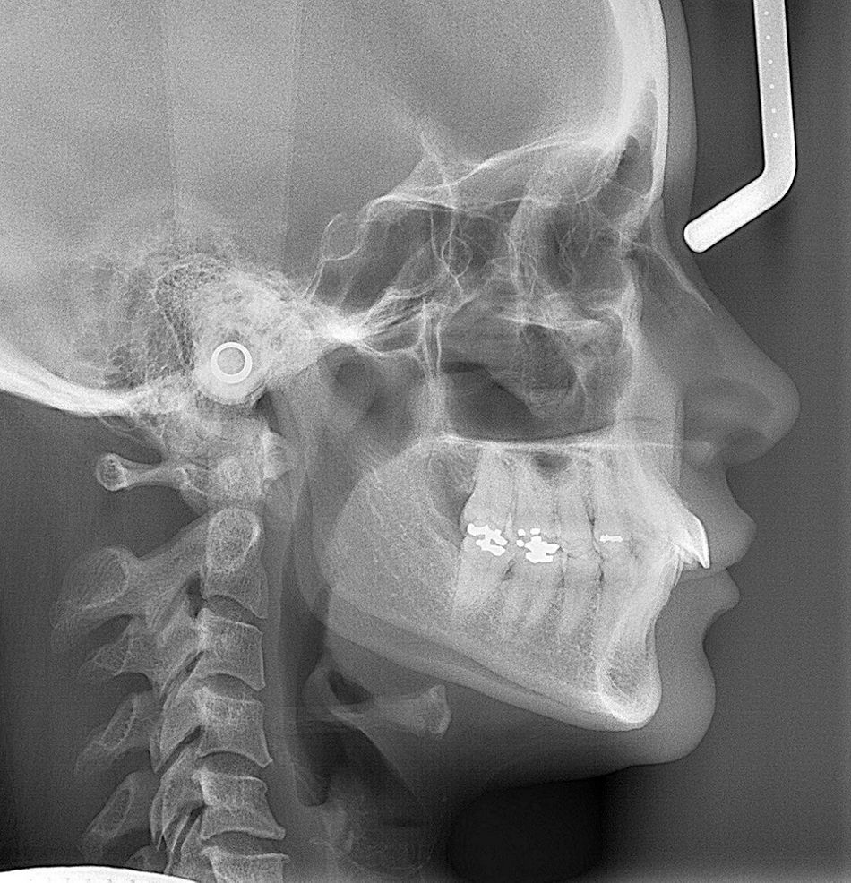
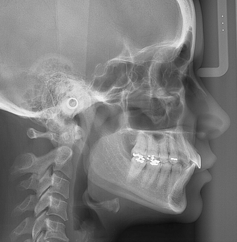
- Fig. 1d: Cephalometric imaging
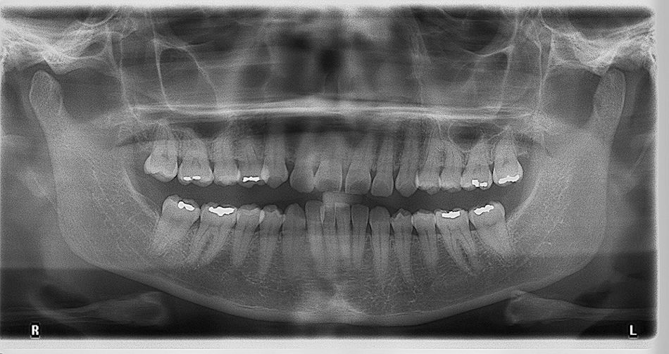
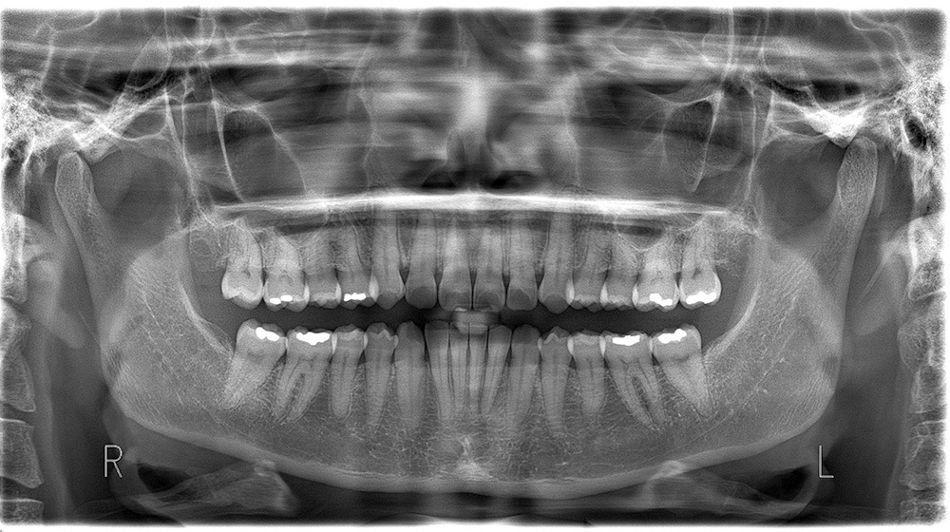
- Fig. 1e: Orthopantomogram
Clinical case study
Anterior deep bites are very often associated with class II occlusion. This is also the case in this 47-year-old patient in whom the severe occlusal interlocking resulted in compression of both mandibular joints and functional complaints. She presented to our orthodontic practice with neck pain, headaches and reciprocal mandibular joint clicking on the right. Thus her primary motivation was functional improvement; although she did also hope for esthetic dental correction with aligner treatment. Esthetically, what bothered her most was the gap between her maxillary incisors. She had already had orthodontic treatment as an adolescent. Manual function analysis showed disc dislocation on the right, with compression of both mandibular joints. Her ability to open her mouth was restricted and, as with closing her mouth, there was deviation.
The extraoral findings showed a convex facial profile with a significantly reduced nasolabial angle (90.8°). There were also impressions of the maxillary teeth in the lower lip.
Cephalometric imaging assessment demonstrated a distal-basal maxillomandibular relationship according to the WITS appraisal (2.6 mm) with slight maxillary prognathism (SNA 86.5°). The dental analysis of the caphalometric imaging showed significant anteinclination of the maxillary incisors (IOK-NL 126.5°) and a manifest anteinclination of the mandibular incisors (IUK-ML 103.6°) with a greatly reduced interincisal angle (IOK-IUK 112.3°). The vertical parameters show a brachycephalic structure. The OPG evaluation showed adult dentition with missing third molars. Root 25 was also extremely curved in a mesial direction. There was moderate generalized horizontal bone loss of about 15% affecting the limbus alveolaris in the upper and lower jaw.
The primary orthodontic diagnosis is an angle class II/1 with enlarged vertical anterior overbite and traumatic bite of the anterior lower jaw into the oral mucosa. Furthermore, the patient had laterognathia to the right due to a posterior forced bite.
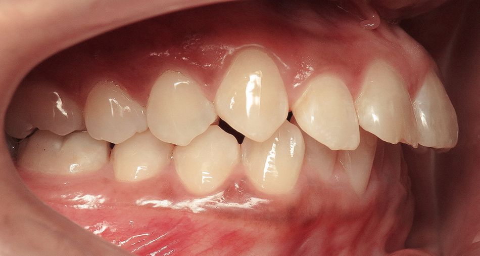
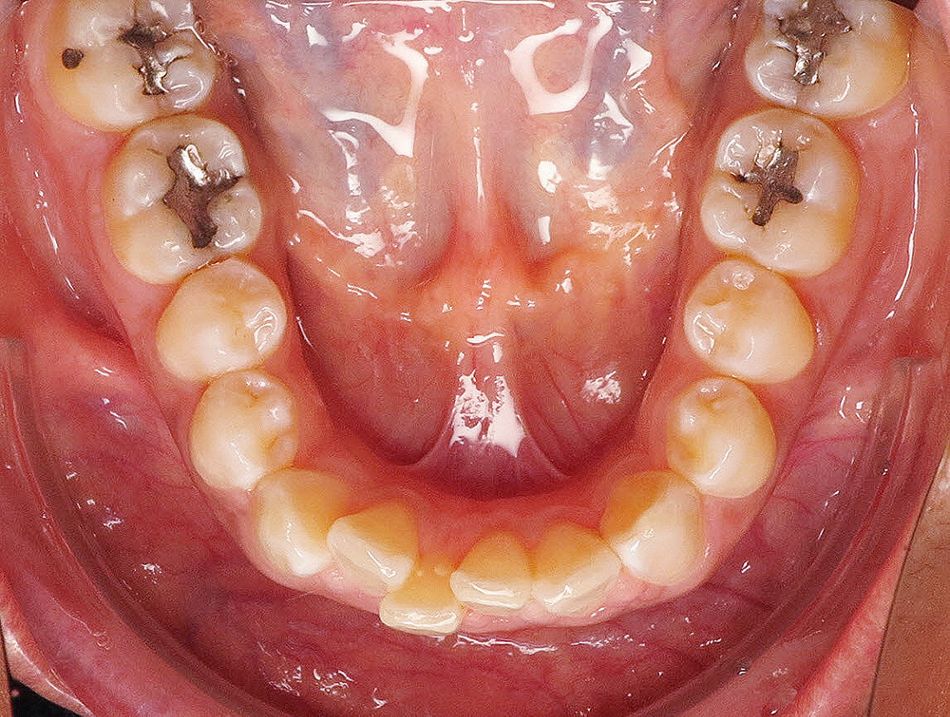
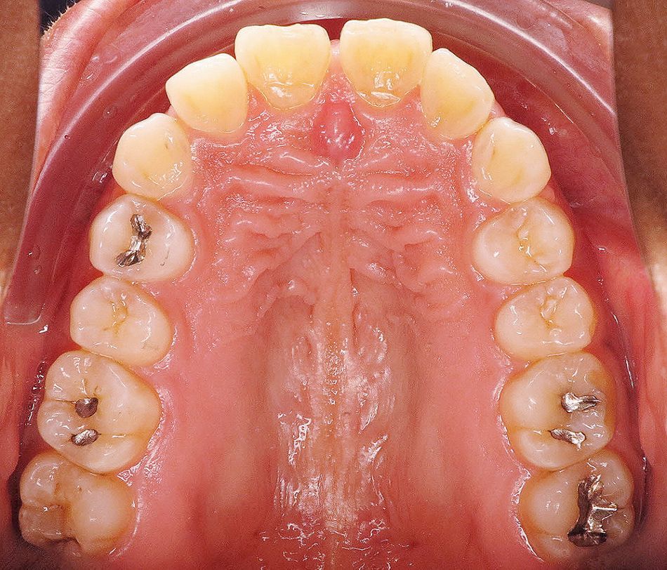
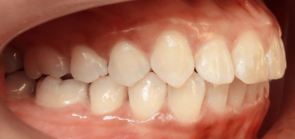
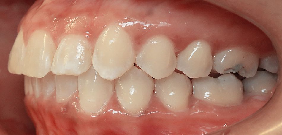
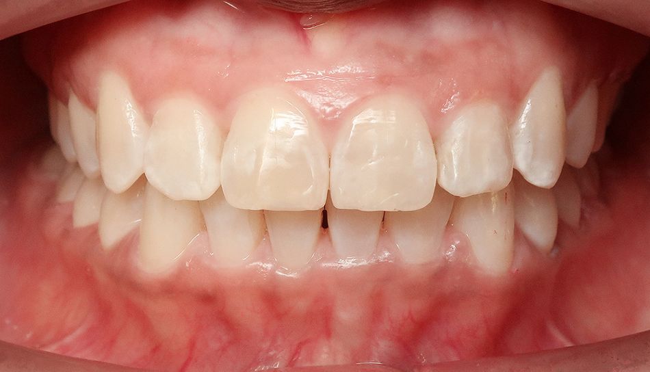
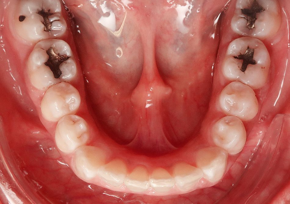
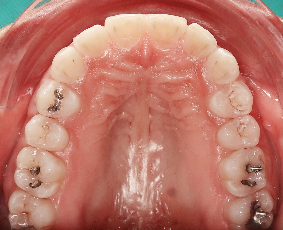
- Fig. 2a: Intraoral images taken at baseline.
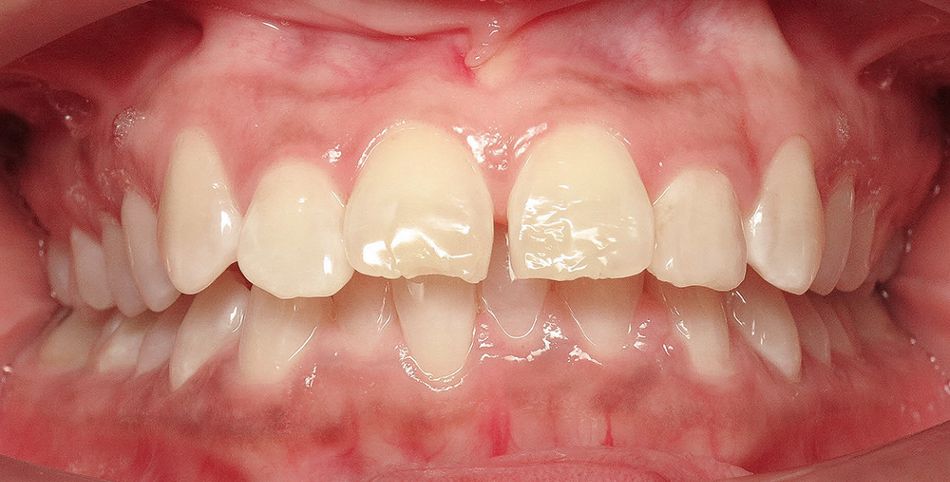
- Fig. 2b: Intraoral images taken at baseline.
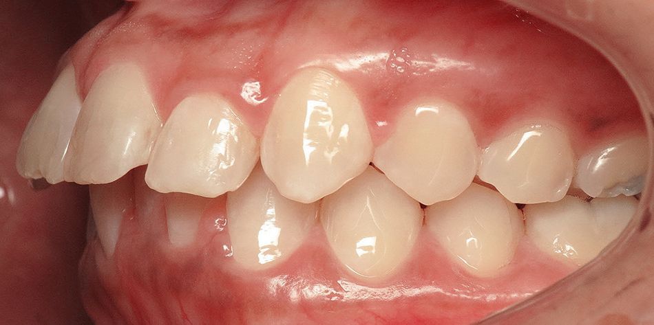
- Fig. 2c: Intraoral images taken at baseline.
- Fig. 2d: Intraoral images taken at baseline.
- Fig. 2e: Intraoral images taken at baseline.
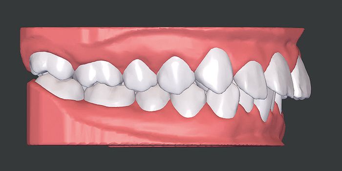
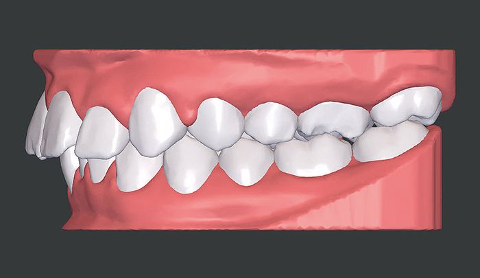
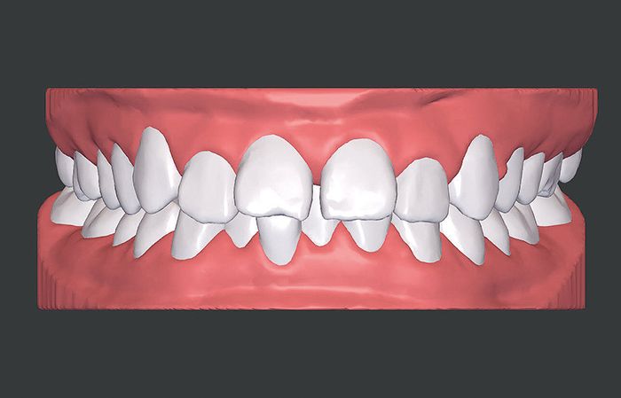
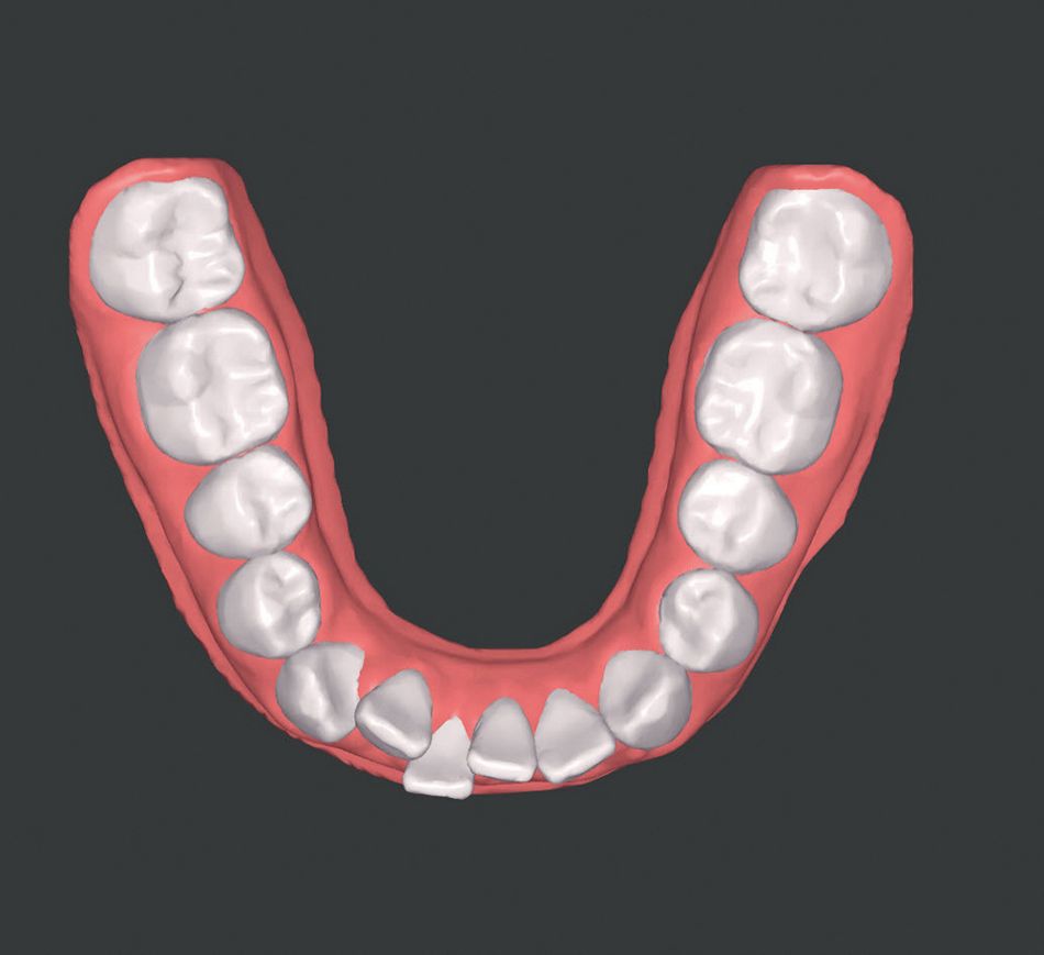
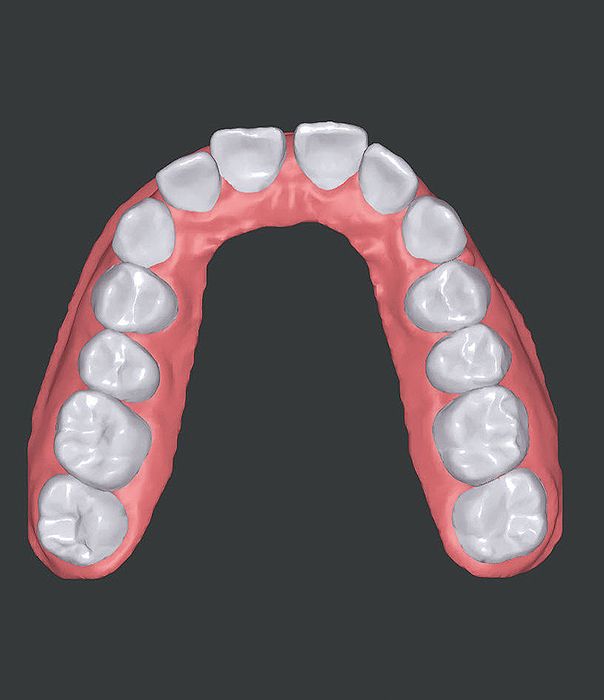
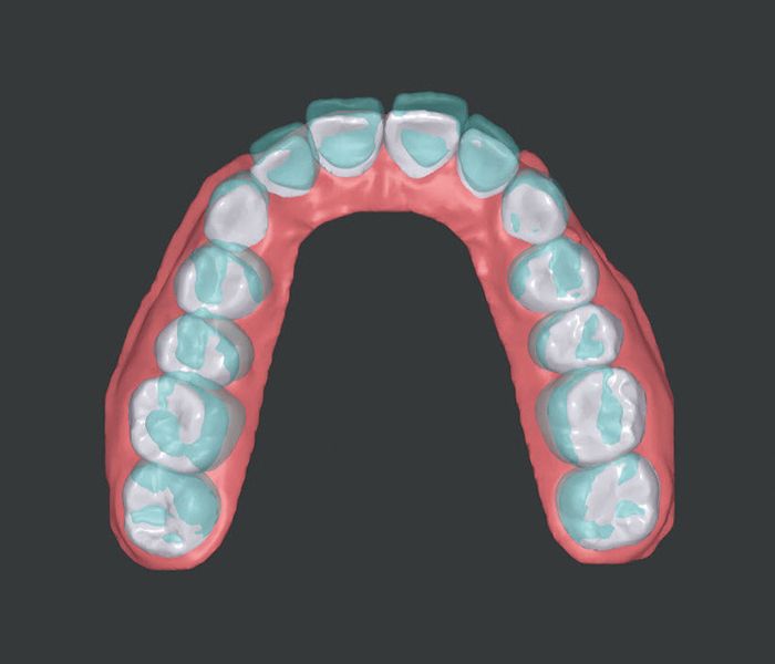
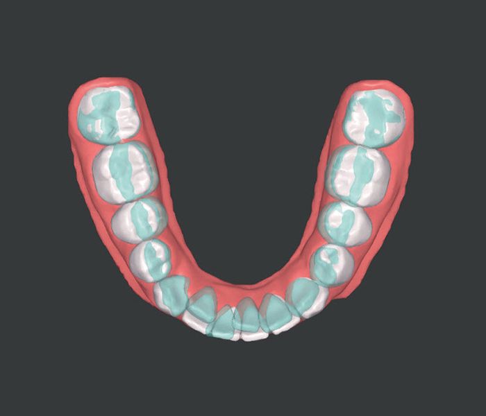
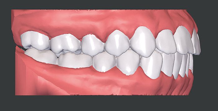
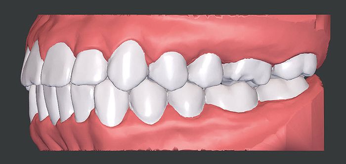
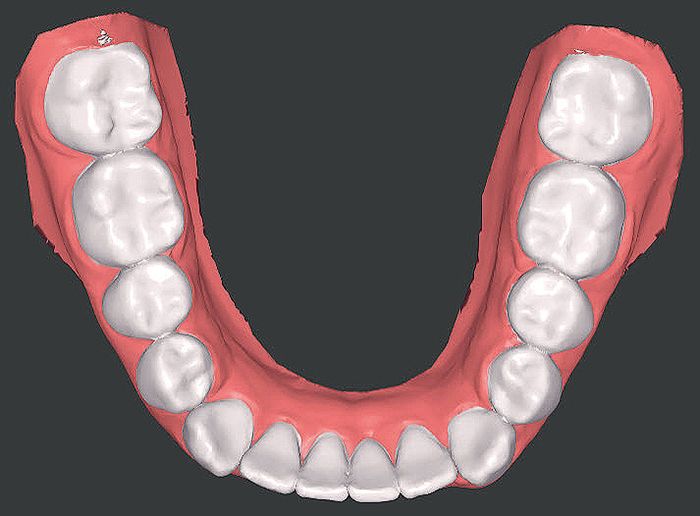
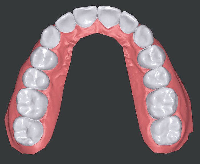
- Fig. 3a: Corresponding baseline situation in ClearPilot® 3D treatment setup.
- Fig. 3b: Corresponding baseline situation in ClearPilot® 3D treatment setup.
- Fig. 3c: Corresponding baseline situation in ClearPilot® 3D treatment setup.
- Fig. 3d: Corresponding baseline situation in ClearPilot® 3D treatment setup.
- Fig. 3e: Corresponding baseline situation in ClearPilot® 3D treatment setup.
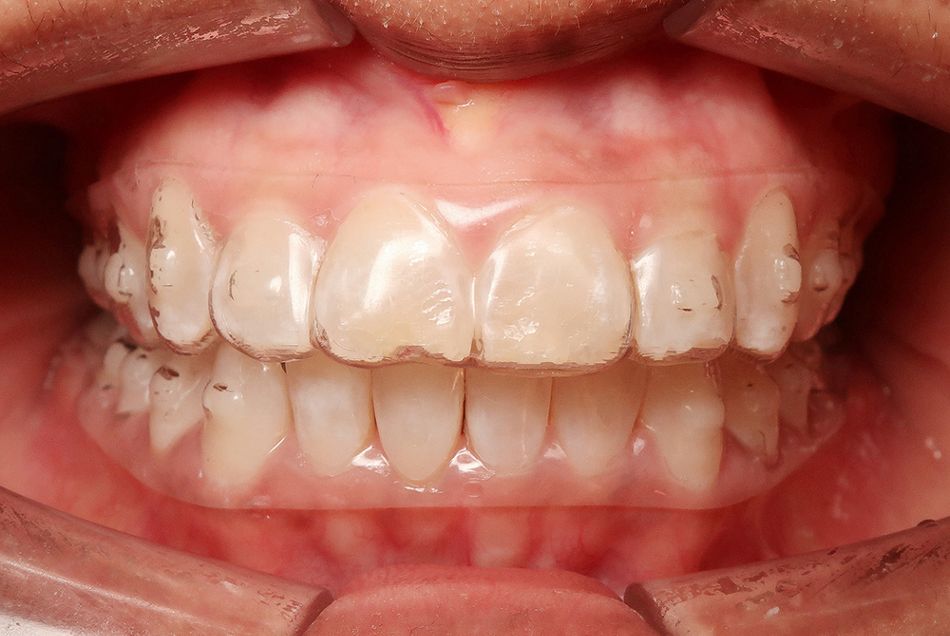
The intraoral findings and the model analysis showed notable anteinclination and elongation of the maxillary incisors as well as midline diastema. Both arches also had transverse narrowness, especially in the anterior region. This was evident in significant mandibular anterior crowding with a steep tilt of teeth 32, 31, 42 and a labial tilt of tooth 41. Abrasions, wear facets, especially in the front, along with isolated areas of gingival recession were observed. On tooth 11 there was an enamel fracture of the incisal edge. Forced laterognathism resulted in a 3 mm midline shift to the right. There was evidence of bilateral distal occlusion with enlarged sagittal overjet (6 mm) and vertical overbite (5 mm). Due to the marked transverse mandibular arch, there was a slight scissor bite on the left.
Following extensive diagnostics and an in-depth discussion with the patient on the risks of treatment and potential alternatives, she opted for treatment with ClearCorrect® aligners. Their high trimline lends these aligners extremely high dimensional stability, making them especially suitable for transverse correction and deep bite treatments. Physiotherapy was also prescribed alongside the approximately 2-year orthodontic treatment.
Digital treatment setup
STL files generated from intraoral scans were uploaded via the ClearCorrect® Doctor Portal. Digital treatment planning was undertaken in collaboration with the ClearCorrect® planning team using the ClearPilot® software.
The main focus was on improving the functional baseline situation by removing the anterior pre-contacts and thereby eliminating the associated posterior forced bite. The treatment also aimed at reducing the vertical anterior overbite and sagittal anterior overjet to achieve occlusion of the anterior teeth.
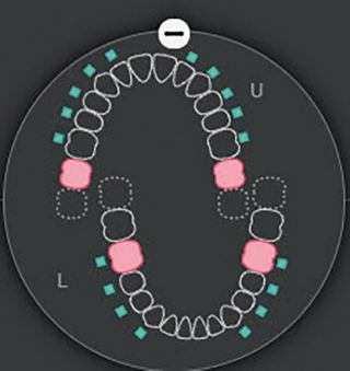
Fig. 4: Sequential distalization demonstrated with the tooth movement chart in ClearPilot®.
Adjustment to achieve class I dentition is also planned with extensive distalization of the maxillary posterior teeth and less extensive distalization of the mandibular posterior teeth. Although the patient had a high trimline with increased retention, a purely sequential distalization was performed to prevent loss of retention¹,². Mandibular molar movement was also combined with tipback and mesial-out rotation to achieve correct axis alignment and contact points. This type of movement also corresponds to the natural movement of the teeth and as such is especially suitable for preserving retention.
The number of aligners – in this case 67 per jaw – was based on the distance the teeth needed to move and segmenting of the individual tooth movements. Great care was taken to ensure that no more than one molar or two premolars/canines were distalized simultaneously per quadrant.
To ensure that retention was observed, a maximum movement of 0.2 mm per aligner was planned³,⁴, although the standard for distalizations with ClearCorrect® is 0.3 mm per aligner. Staging and thus also the speed at which the teeth move were adjusted by appropriate instructions in the prescription.
Expansion in the mandible and intrusion of the anterior mandible were planned in segments. Intrusion of the mandible and anterior maxilla, especially teeth 12 and 22, were overcorrected in the 3D treatment planning with ClearPilot®.
When retrusion of the anterior teeth was scheduled towards the end of treatment, care was taken to implement a root torque to avoid further gingival recession and to prevent an excessively steep maxillary anterior at the end of the anterior retrusion.
Attachment planning
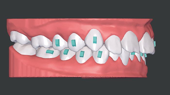
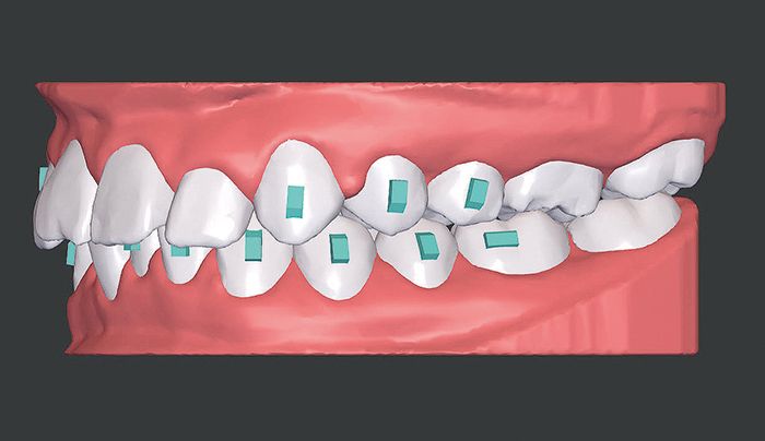
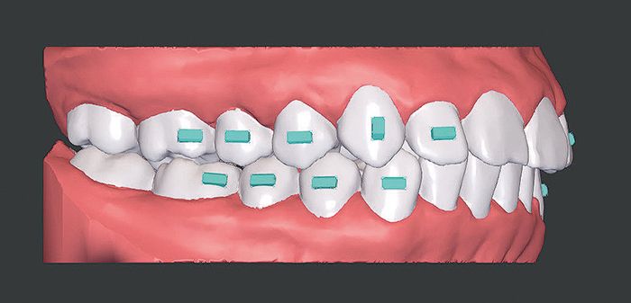
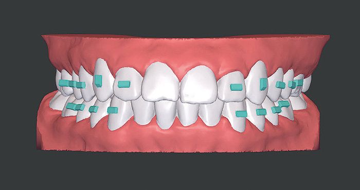
Vertical rectangular attachments were planned for the mandibular canines and premolars to make intrusion of the anterior teeth more predictable. The horizontal rectangular attachments on the 1st mandibular molars were used to compensate for the retention forces generated by Class II elastics. At the same time, vertical rectangular attachments were planned on the maxillary canines and premolars to ensure maxillary incisor intrusion.
- Fig. 5a: Attachment planning of the ClearCorrect® treatment, attachment placement from aligner level 3.
- Fig. 5b: Attachment planning of the ClearCorrect® treatment, attachment placement from aligner level 3.
- Fig. 5c: Attachment planning of the ClearCorrect® treatment, attachment placement from aligner level 3.
Treatment
The first two aligners were – as planned in the ClearPilot® software – worn without attachments to allow the patient to get used to wearing them. Before the third aligner, two transfer trays (templates) were used to position the attachments onto the teeth with packable composite. Aligner replacement was initially scheduled every 14 days; this interval was set to be shortened over the course of treatment.
The initial 14-day interval to change the aligners is recommended as smaller orthodontic tooth movement is expected at the start of treatment, since the reconstruction processes controlled by osteoblasts and osteoclasts only occur after about 2-4 weeks⁵,⁶. Thus a shorter interval at the start of treatment is not recommended.
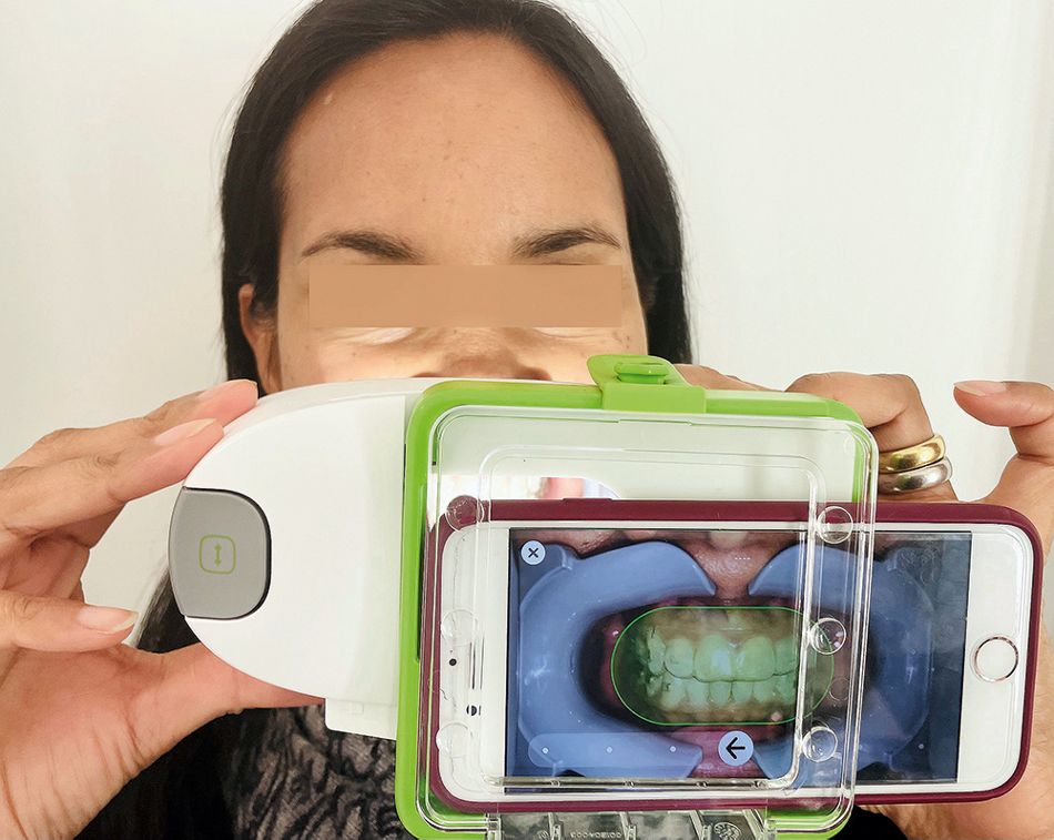
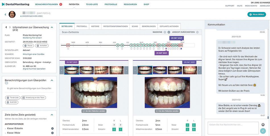
In addition to the scheduled recall appointments, the patient had supplementary digital treatment check-ups using Dental Monitoring™. Artificial intelligence was deployed to evaluate intraoral photos taken with the smartphone, allowing the aligner fit to also be monitored remotely throughout treatment.
The length of time each aligner was worn was adjusted to the actual clinical situation. As the patient herself considered the aligner fit to be good, and given that the fit could be remotely monitored by the AI after each scan, the interval at which the aligners were changed could be reduced to 7-10 days (dynamic aligner change). The technology also allowed the quality level to be checked continuously and offered the opportunity to act early rather than wait until the next scheduled check-up. The information on the clinical objectives with Dental MonitoringTM saved time during the treatment and meant that appointments were only arranged if there were clinically relevant events. The fact that the patient was not scheduled for intraproximal reduction (IPR) also helped.
- Fig. 6a: Patient taking the Dental Monitoring TM scan using the Scan Box.
- Fig. 6b: Dental Monitoring TM Portal.
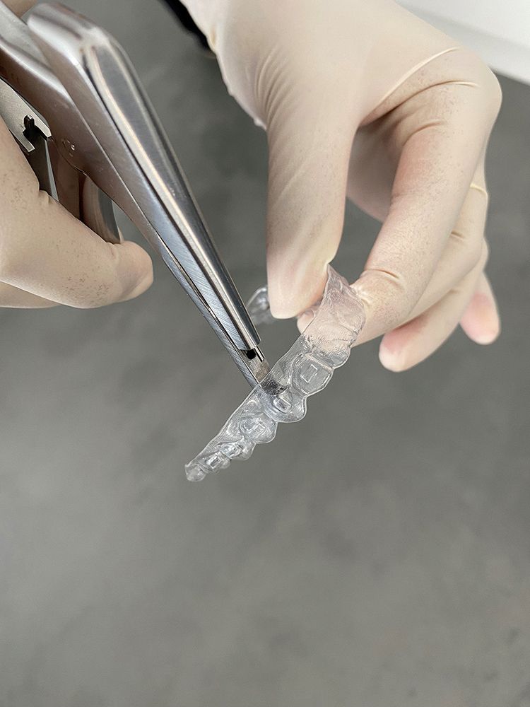
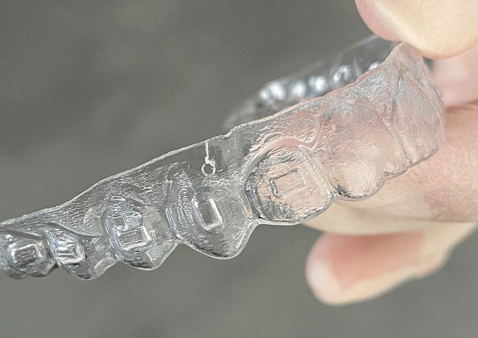
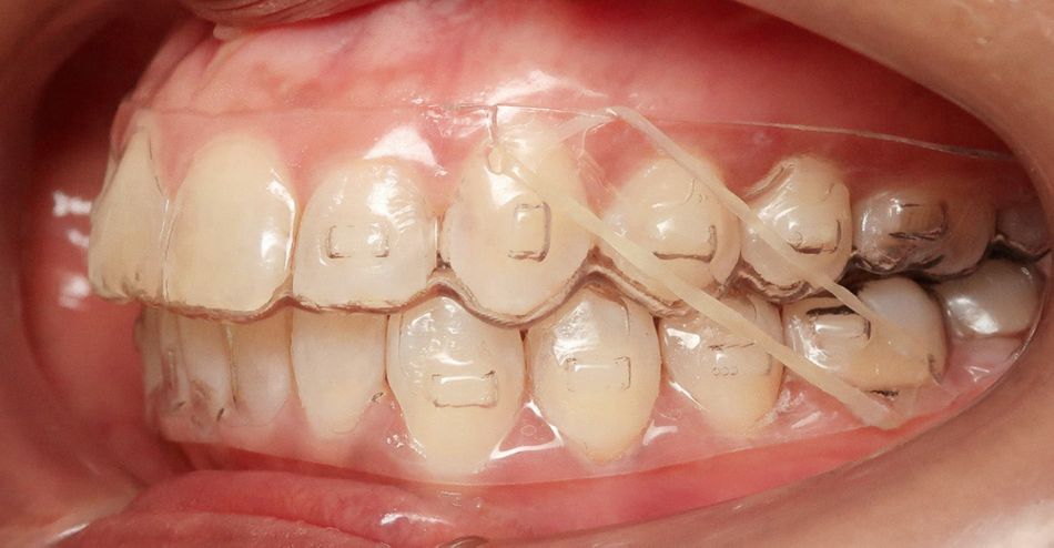
The patient wore bilateral class II elastics at night (4 ½ oz./6.4 mm) for additional retention support for sequential distalization in the maxilla, albeit only once the slight distalization in the mandible was concluded. As it was not possible at the time of treatment to order cut outs for buttons, the aligners were processed with the aligner punch slot machine from Hammacher. This was carried out mesial of the upper canines and distal of the mandibular first molars. In contrast to cutting these with scissors or a ligature cutter, this procedure prevented damage to the aligner and allowed the elastics to be securely attached to the aligners.
- Figs. 7a: Aligner punch slot machine from Hammacher in use
- Figs. 7b: Aligner punch slot machine from Hammacher in use
- Fig. 7c: Class II elastics (4 ½ oz. /6.4 mm) on the left
The patient came for treatment check-ups at different intervals ranging from 8 to 20 weeks. This was possible because the complete treatment was conducted without approximal enamel reduction and because the patient had regular supplementary digital treatment check-ups with Dental Monitoring™.
Interim findings
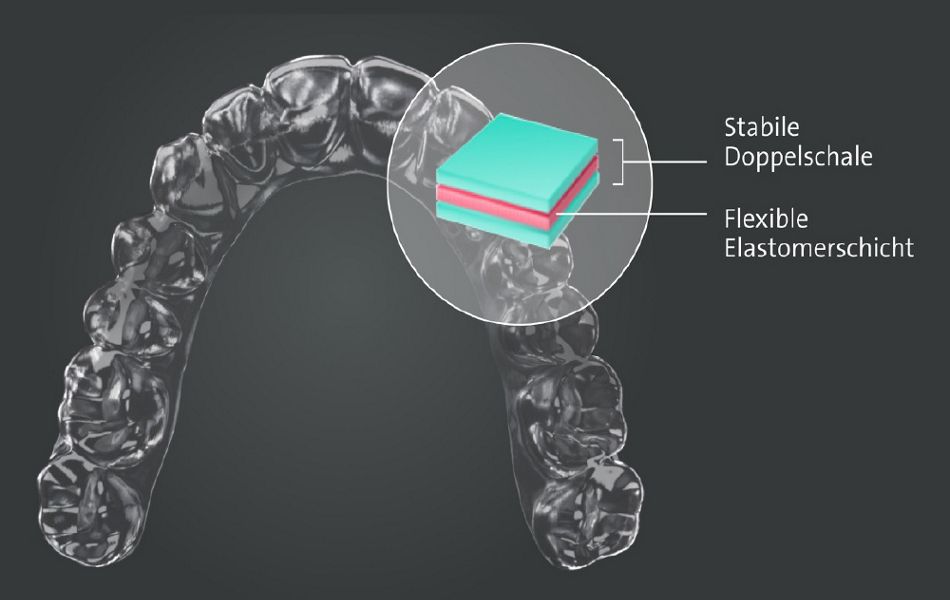
To achieve the desired treatment objective, another alignment order had to be placed. The patient was informed at this time about the pending material switch at ClearCorrect®. The new three-layer ClearQuartz® material, which according to the manufacturer consists of two hard outer shells sandwiching a flexible elastomer layer is designed to increase the predictability of translation and rotation movements. Although this resulted in a retention phase of 20 weeks, the patient opted to switch to the new material for what is known as the refinement phase (revision).
- Fig. 8: The 3-layer ClearQuartz® aligner material is a third-generation development consisting of an internal elastomer layer sandwiched between resistant outer layers with low porosity.
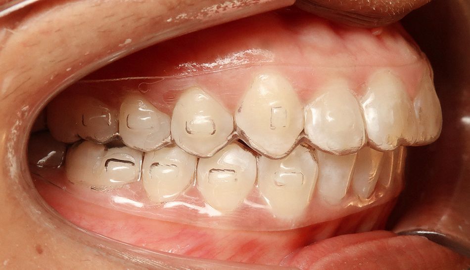
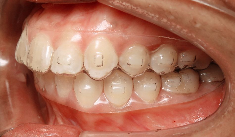
The interim findings in the refinement planning showed that the bite was already significantly raised and that the occlusion of the posterior teeth had improved. The aligner fit with the specific high trimline can be described as very good.
- Fig. 8a: Interim findings for aligner 67. Baseline before refinement. The high trimline and snug aligner fit are clearly evident.
- Fig. 8a: Interim findings for aligner 67. Baseline before refinement. The high trimline and snug aligner fit are clearly evident.
- Fig. 8a: Interim findings for aligner 67. Baseline before refinement. The high trimline and snug aligner fit are clearly evident.
- Fig. 8b: Digital presentation at the start of refinement.
- Fig. 8b: Digital presentation at the start of refinement.
- Fig. 8b: Digital presentation at the start of refinement.
- Figs. 9a: Superimposition tool maxilla and mandible in the ClearPilot® 3D treatment setup step 1-67.
- Figs. 9b: Superimposition tool maxilla and mandible in the ClearPilot® 3D treatment setup step 1-67.

Want to stay up to date?
youTooth.com is THE PLACE TO BE IN DENTISTRY – subscribe now and receive our monthly newsletter on top hot topics from the world of modern dentistry.
Treatment completion
- Fig. 10a: Final images; final extraoral patient photographs, front and profile (a-c), caphalometric imaging of posterior teeth (d) and orthopantomogram (e).
- Fig. 10b: Final images; final extraoral patient photographs, front and profile (a-c), caphalometric imaging of posterior teeth (d) and orthopantomogram (e).
- Fig. 10c: Final images; final extraoral patient photographs, front and profile (a-c), caphalometric imaging of posterior teeth (d) and orthopantomogram (e).
- Fig. 10d: Final images; final extraoral patient photographs, front and profile (a-c), caphalometric imaging of posterior teeth (d) and orthopantomogram (e).
- Fig. 10e: Final images; final extraoral patient photographs, front and profile (a-c), caphalometric imaging of posterior teeth (d) and orthopantomogram (e).
- Fig. 11a: Intraoral images of the final situation.
- Fig. 11b: Intraoral images of the final situation.
- Fig. 11c: Intraoral images of the final situation.
- Fig. 11d: Intraoral images of the final situation.
- Fig. 11e: Intraoral images of the final situation.
- Fig. 12a: Corresponding final situation in the ClearPilot® 3D treatment planning.
- Fig. 12b: Corresponding final situation in the ClearPilot® 3D treatment planning.
- Fig. 12c: Corresponding final situation in the ClearPilot® 3D treatment planning.
- Fig. 12d: Corresponding final situation in the ClearPilot® 3D treatment planning.
- Fig. 12e: Corresponding final situation in the ClearPilot® 3D treatment planning.
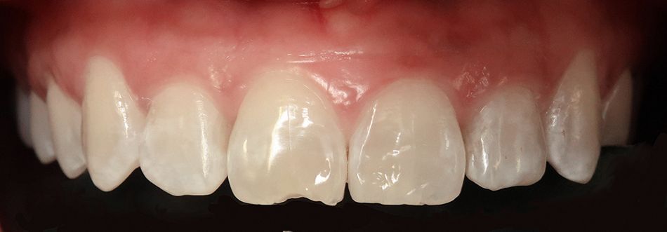
After refinement is completed, bilateral neutral occlusion was achieved in the first molar region. The nasolabial angle was improved with a significantly reduced overjet and overbite with unstrained lip closure. Since the patient’s incisors were previously crowded and she did not undergo interdental enamel reduction, a black triangle persisted in the region of teeth 31/41, which could be rectified with a slight composite build-up. The abfractions on the cutting edge on tooth 11 were restored with composite.
- Figs. 13a: Build-up of the cutting edge on tooth 11.
- Figs. 13b: Build-up of the cutting edge on tooth 11.
Overall, a significant functional improvement was achieved, resolving the headaches and neck pain as well as the jaw clicking the patient had been experiencing. Her mouth opening was also significantly improved. Retention in the mandible was achieved with a six-point retainer (5-stranded .0155 lingual retainer wire 24K gold-plated, KFO24 GmbH in Cologne) and additional Hawley retainer in the maxilla and mandible.
Discussion
Digital treatment setup was very difficult, since there were tooth movements in all three planes and because the Tooth Editing function was not available when planning was carried out. The final tooth position could only be adjusted by communicating with ClearCorrect® technicians by text, using a function available in the software.
The advantage of the ClearCorrect®-specific high trimline and the associated form stability certainly meant that the planned transversal correction could be clinically fully implemented. The bite could also be successfully raised, although BiteRamps were not available.
As well as intrusion of the anterior arch and improved posterior occlusion with sequential distalization of the posterior teeth, a retrusive alignment of the anterior teeth was also achieved. This made lip closure more comfortable and slightly improved the facial profile.
The patient herself reported a significant difference between the material used at the start of treatment and the ClearQuartz® material of the refinement. With the tooth movements at roughly equal increments, the patient found the fit to be better and the force to be more evenly distributed, one indication of which was less initial tension when she started wearing a new aligner. Integrating Dental Monitoring™ into the treatment processes meant that there were far fewer in-person maintenance visits to our practice, an option that has considerable benefits in terms of time and cost both for working patients as well as school pupils.
Conclusion
The ClearCorrect® treatment system appears not only to be suitable for simple dental alignment corrections thanks to its material properties and the shape of the aligners, but can also demonstrate its strength in pronounced three-dimensional deviations of the arch shape.
Based on the positive experience, we are considering using Dental Monitoring™ in all patients in our practice as a matter of routine. The associated compliance benefits and the easier hygiene routines are significant advantages.
Outlook
At the start of 2022, ClearCorrect® is launching a new version of ClearPilot®. This version gives dentists full 3D control over tooth movements, allowing them to move teeth independently to the final position using a 3D dialog tool while simultaneously achieving the desired occlusal contacts. The occlusion can also be checked in the 3D model with transparent arches. The software also offers a multi-view (arches viewed from different perspectives) and in-App navigation to previous planning versions.
- Fig. 14: 3D control within the ClearPilot® treatment setup using individual tooth movements
Photographs: © Dr. Jörg Schwarze / © Straumann
© First publication: Oemus Media AG, KN, Kieferorthopädie Nachrichten, 2021; No. 12, page 1, 6-10.
Literature
- Ravera S., Castroflorio T. et al. Maxillary molar distalization with aligners in adult patients: a multicenter retrospective study. Prog Orthod. 2016;17:12.
- Caruso S., Nota A. et al. Impact of molar teeth distalization with clear aligners on occlusal vertical dimension: a retrospective study. BMC Oral Health. 2019 Aug 13;19(1):182.
- Simon M., Keilig L., Schwarze J. et al. Forces and moments generated by removable thermoplastic aligners: incisor torque, premolar derotation, and molar distalization. Am J Orthod Dentofacial Orthop. 2014; 146(4):411.
- Simon M, Keilig L, Schwarze J et al. Treatment outcome and efficacy of an aligner technique--regarding incisor torque, premolar derotation and molar distalization. BMC Oral Health. 2014; 14:68.
- Arrifin S., Yamamoto Z. et al. Cellular and molecular changes in orthodontic tooth movement. ScientificWorldJournal. 2011; 11: 1788–1803.
- Diedrich, Peter (2000). Kieferorthopädie II. Grundlagen kieferorthopädischen Gewebeumbaus. Urban & Fischer: München.
- Guarneri M, Gracco A, Farina A, Schwarze J. Attachment of intermaxillary elastics to thermoformed aligners. J Clin Orthod. 2009 Jan;43(1): 34-7.


