#Esthetics 03. Jan 2024

Peri-implantitis treatment with GalvoSurge® in the esthetic zone: One approach for biofilm removal in two patients

by Alberto Monje, Spain

Introduction

Peri-implantitis is a pathological condition occurring in tissues around dental implants and characterized by inflammation in the peri-implant connective tissue and progressive loss of supporting bone1.

Particularly, peri-implantitis poses a significant clinical challenge in the esthetic zone due to its potential to cause mucosal recession. This condition can lead to visible changes in the appearance of the implant site, impacting overall esthetics. Managing peri-implantitis in such areas requires careful consideration and tailored strategies to prevent or address mucosal recession and preserve the esthetic outcome2.

In treating peri-implantitis, the goal is to reduce inflammation in soft tissues and stop further bone loss. Since nonsurgical methods have their limitations in achieving this goal, surgery is often recommended, and different techniques have been proposed based on factors like bone configuration and soft tissue characteristics3.

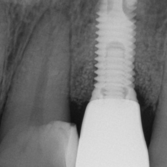
Before initiating any regenerative treatment, it is essential to eliminate biofilm around the dental implant. In recent years, several modalities and innovations have emerged for this purpose. One example is the GalvoSurge®️ Dental Implant Cleaning System, a biofilm-removal technology based on electrolytic cleaning. In just two minutes, GalvoSurge® removes bacterial biofilm from dental implants affected by peri-implantitis, setting the stage for bone grafting procedures.

This report unfolds a narrative of innovation in biofilm control and precision in bone regeneration, showcasing the potential of GalvoSurge®️ in treating peri-implantitis in two patients with affected peri-implant tissues within the esthetic zone4.

Keys for success

  • The configuration of the peri-implant defect may play a pivotal role in achieving successful reconstructive outcomes, leading to disease resolution and marginal bone level gain.
  • Narrow defect angle (<40 degrees) may further favor the reconstructive outcome.
  • In cases where the implant is within the bony housing, the recommended approach involves surface decontamination followed by regeneration. 
  • In cases with a 2-wall defect configuration, the use of a barrier membrane is preferable. Meanwhile, in scenarios involving a well-contained defect configuration, barrier membrane might be optional.
  • For enhanced esthetic outcomes, soft tissue grafting using a connective tissue graft is advisable.

Patient No. 1
Initial situation

A patient came to the clinic complaining of discomfort, bleeding and pus discharge in the anterior esthetic area (Fig. 1.1). The intraoral examination showed deep pocketing around implant #22, and radiographic analysis indicated moderate bone loss (Fig. 1.2).

Treatment planning

The treatment plan was discussed with the patient, and a mucoperiosteal flap with mechanical instrumentation combined with GalvoSurge® was decided. Guided bone regeneration was planned to address the bone defects.

Surgical procedure

A mucoperiosteal flap was elevated by means of intrasulcular incisions. A peri-implantitis-related lesion was meticulously removed from the implant using a site-specific curette. A moderate 2-wall peri-implantitis-related bone defect (class Ib) in the esthetic area was observed (Figs. 1.3–1.4). The configuration and depth of the defect are crucial factors in evaluating the feasibility of a therapeutic approach. The implant, situated within the bony housing, was deemed suitable for reconstructive therapy. Mechanical surface decontamination was performed using a titanium brush (Fig. 1.5). An electrolytic approach, utilizing the GalvoSurge® Dental Implant Cleaning System, was employed to decontaminate the surface and establish an aseptic environment (Fig. 1.6).

To support bone grafting, a resorbable barrier membrane was utilized (Fig. 1.7). A 1:1 combination of deproteinized bovine bone mineral and autologous bone chips obtained from adjacent surgical sites was applied to graft the bone defect (Fig. 1.8). A porcine collagen membrane was secured with subperiosteal sutures (Fig. 1.9). Further stabilization involved the use of a subepithelial connective tissue graft to attain mucosal stability and minimize recession (Fig. 1.10).

Treatment outcomes at 20-month follow-up

At the 20-month follow-up, clinical assessment revealed complete resolution of the disease (Fig. 1.11). Reduction of probing pocket depth and bleeding on probing was noted. Substantial radiographic bone level gain and stability were observed during the same period (Fig. 1.12). The patient was pleased with the esthetic outcome.

Patient No. 2
Initial situation

During the patient's annual check-up at the referral practice, it was noted that implant# 22 exhibited signs of bleeding on probing and deep pocketing (Fig. 2.1). A radiograph was taken, and moderate bone loss was observed (Fig. 2.2).

Treatment planning

Surgical plaque removal with guided bone regeneration was planned. The surgical phase plays a crucial role in achieving effective plaque removal and has demonstrated efficacy, particularly in cases with moderate and severe pocket depths.

Surgical procedure

After performing the flap, the crown of #22 was removed to facilitate access and mechanical instrumentation. At this point, it was possible to observe a moderate 3-wall peri-implantitis-related bone defect (class Ib) in the esthetic area. The peri-implantitis-related lesion was removed using a site-specific curette (Fig. 2.3). Subsequently, a titanium brush was employed to remove calculus from the implant surface, followed by the electrolytic approach using GalvoSurge® to remove bacterial biofilm and achieve surface decontamination.

To support bone grafting, a resorbable barrier membrane was utilized. Autologous bone chips were harvested from adjacent surgical sites and were subsequently mixed with deproteinized bovine bone mineral to completely fill the defect for reconstructing the lost peri-implant tissues. The collagen membrane was secured through subperiosteal sutures. Additionally, a subepithelial connective tissue graft was employed and stabilized to attain mucosal stability, minimizing recession. The wound was closed in a tension-free manner to promote transmucosal healing (Figs. 2.5-2.9).

Treatment outcomes at 15-month follow-up

At the 15-month follow-up, clinical assessment revealed complete resolution of the disease. The esthetic outcome demonstrated harmony with limited mucosal recession. Additionally, substantial radiographic bone level gain and stability were observed during the same period (Figs. 2.10-2.12).

Author’s message

The management of peri-implantitis from the esthetic standpoint is challenging, as it entails the achievement and maintenance of a healthy ecosystem, minimizing esthetic distortion, and producing a harmonious soft tissue profile. The use of GalvoSurge® for surface decontamination, combined with bone reconstruction simultaneous to soft tissue grafting by means of a sub-epithelial connective tissue graft, demonstrated safety and efficacy in attaining success.

References:

  1. Schwarz F, Derks J, Monje A, Wang HL. Peri-implantitis. J Periodontol. 2018 Jun;89 Suppl 1:S267-S290. doi: 10.1002/JPER.16-0350. PMID: 29926957.
  2. Ramanauskaite A, Obreja K, Sader R, Becker J, Schwarz F. Assessment of soft and hard tissue dimensions following different treatment approaches of ligature-induced peri-implantitis defects. Clin Oral Implants Res. 2021; 32: 394-400.
  3. Monje A, Amerio E, Cha JK, Kotsakis G, Pons R, Renvert S, Sanz-Martin I, Schwarz F, Sculean A, Stavropoulos A, Tarnow D, Wang HL. Strategies for implant surface decontamination in peri-implantitis therapy. Int J Oral Implantol (Berl). 2022 Sep 9;15(3):213-248. PMID: 36082658.
  4. Monje A, Mesquita PF. Clinical considerations in the surgical management of peri-implantitis lesions in the esthetic zone. J Esthet Restor Dent. 2023 Apr;35(3):457-466. doi: 10.1111/jerd.12921. Epub 2022 May 6. PMID: 35521693.