#Esthetics 21. Mar 2017

Straumann® Bone Level Tapered Implant and augmentation with cerabone®, Jason® membrane and Emdogain® in daily practice

A clinical case report by Michael Kristensen, Denmark

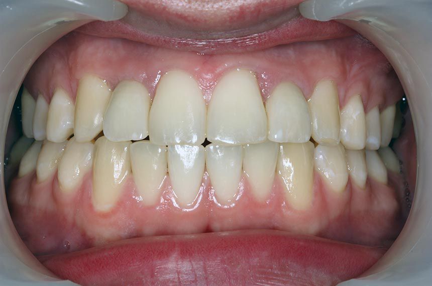
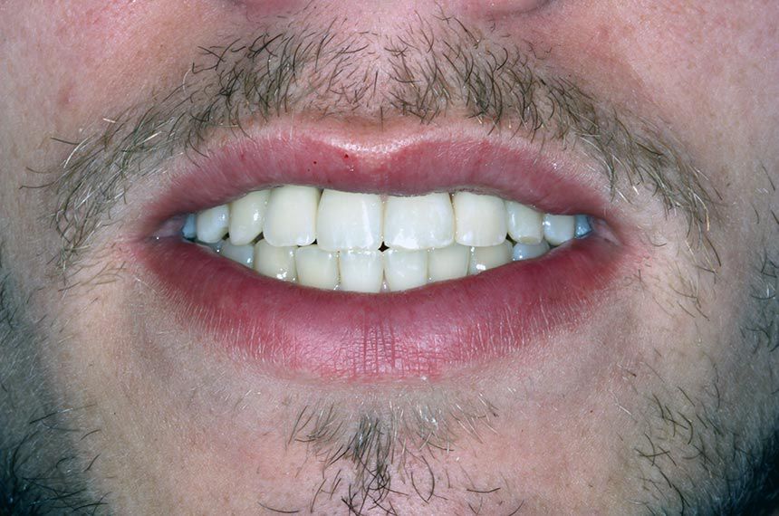
“Straumann® BLT implants in combination with botiss biomaterials (cerabone® and Jason® membrane) and Straumann Emdogain® made this challenging case uncomplicated, with only minimal patient discomfort after surgery and a very good esthetic result.” Dr. Michael Kristensen
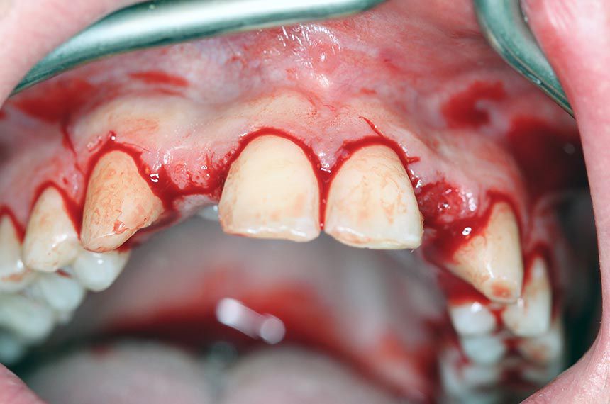
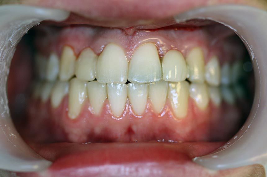
Initial situation

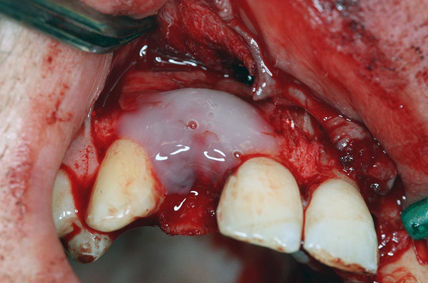
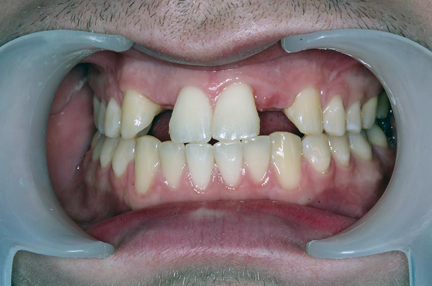
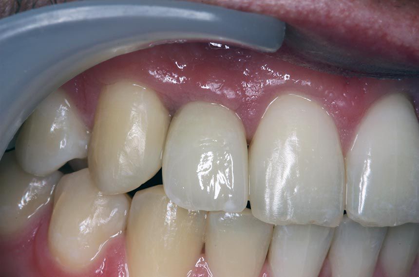
The patient, a healthy 22-year-old male, has agenesis of 12 and 22, which had previously been treated with Maryland bridges. (Fig. 1) He was referred to the implant clinic by a colleague for esthetic reasons.
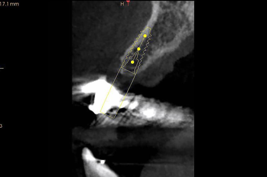
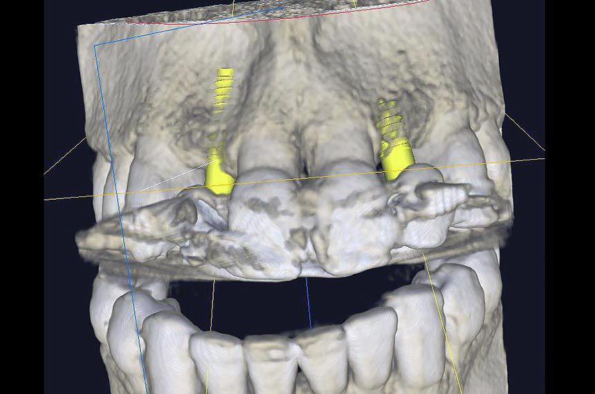
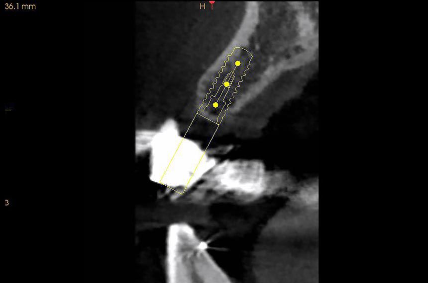
Treatment planning

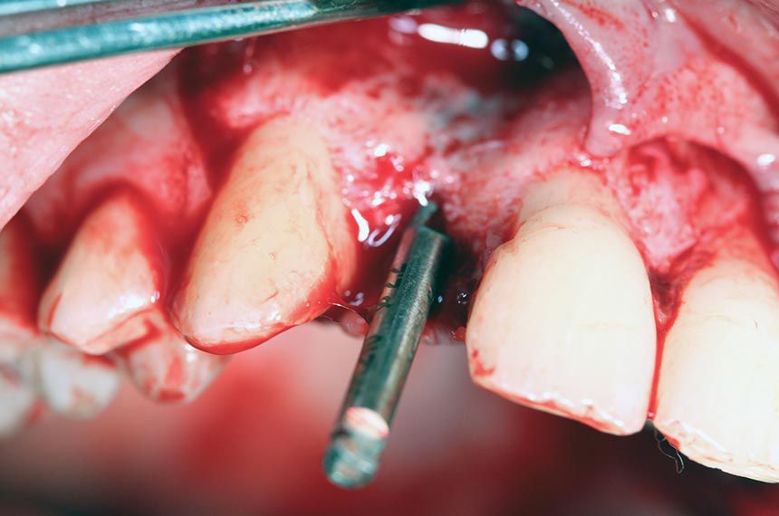
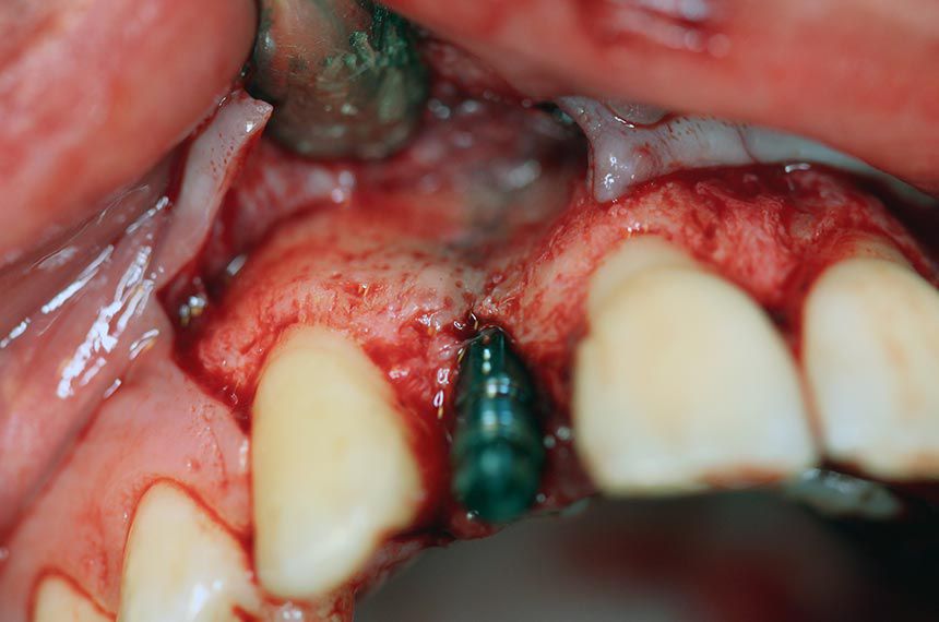
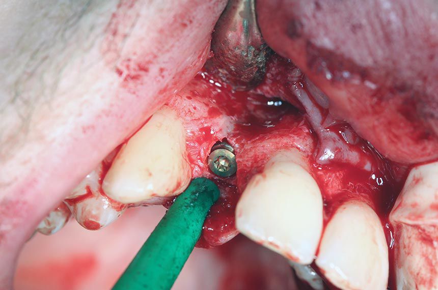
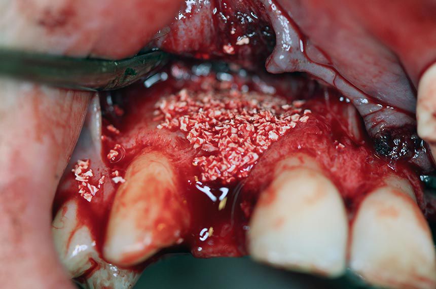
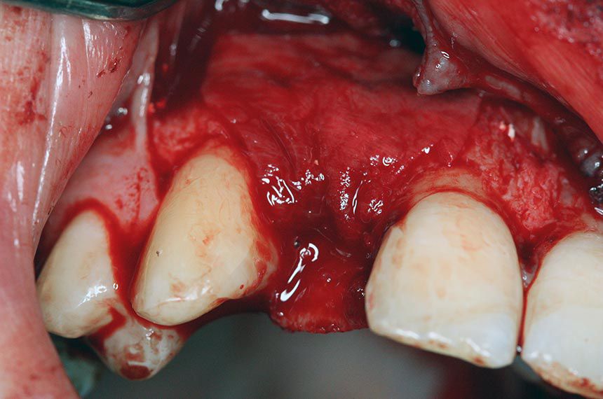
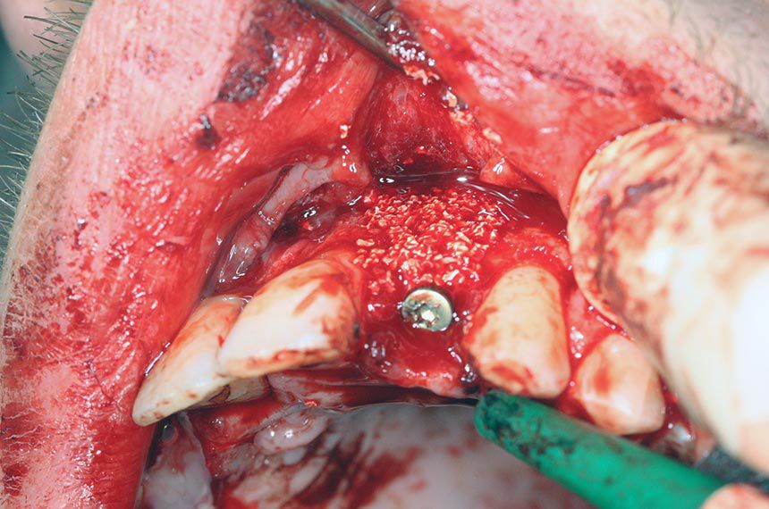
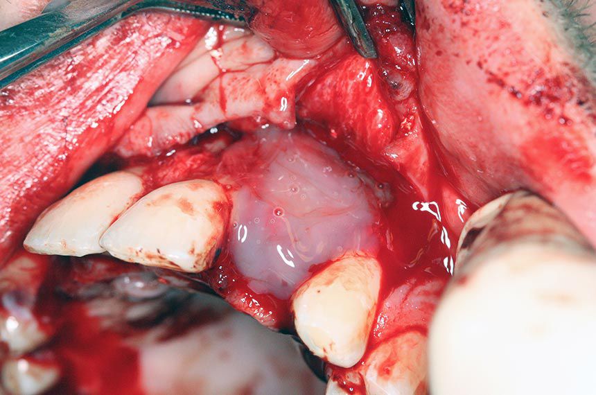
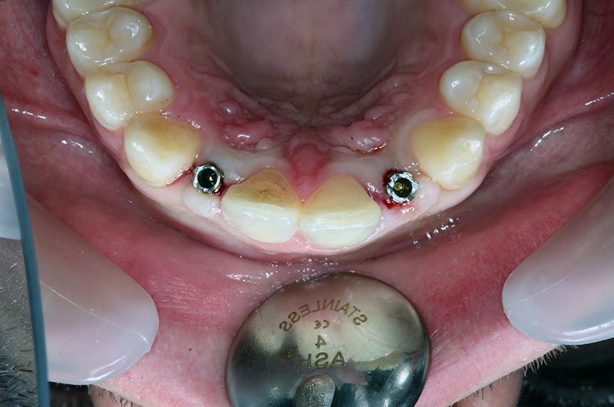
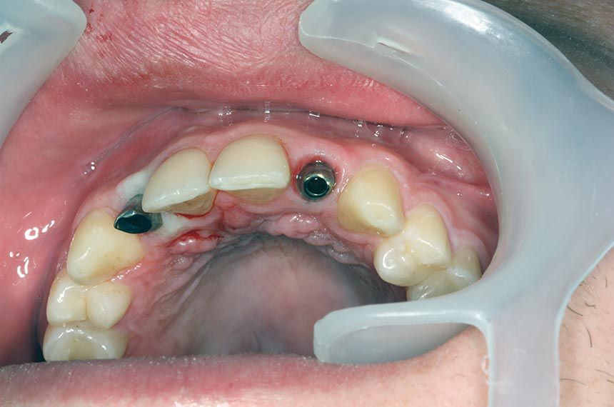
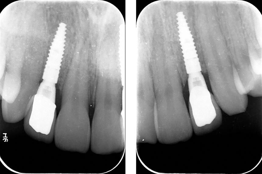
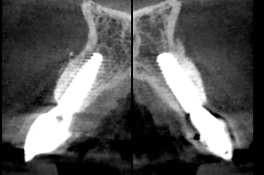
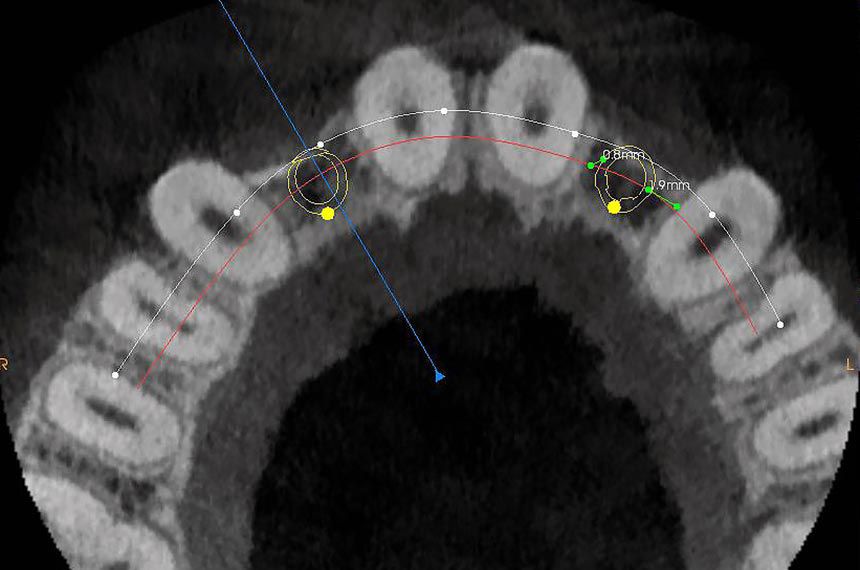
CBCT scans (Figs. 2-5) showed limited space between the roots, but this was sufficient for 3.3 mm Straumann® Bone Level Tapered (BLT) Implants. The ridge was very thin (as in all agenesis patients) and needed horizontal augmentation in order to have at least 1 mm of bone on the buccal side. Since the bone volume was sufficient for primary stability of the implants, the horizontal augmentation was done together with insertion of the implants.
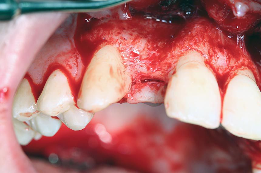
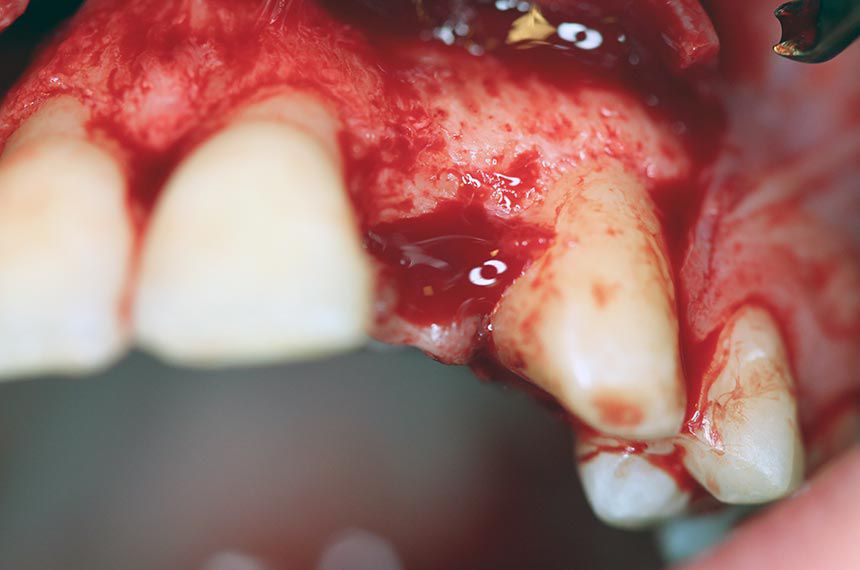
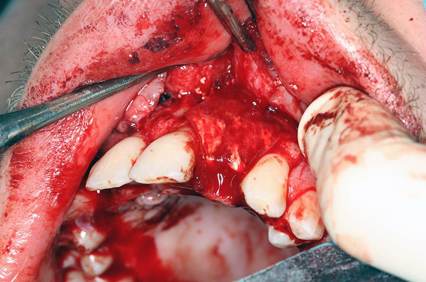
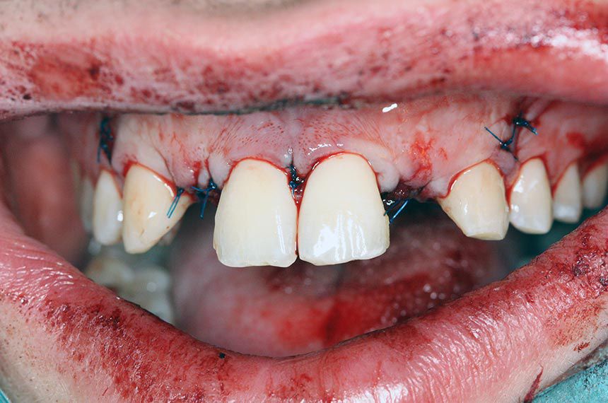
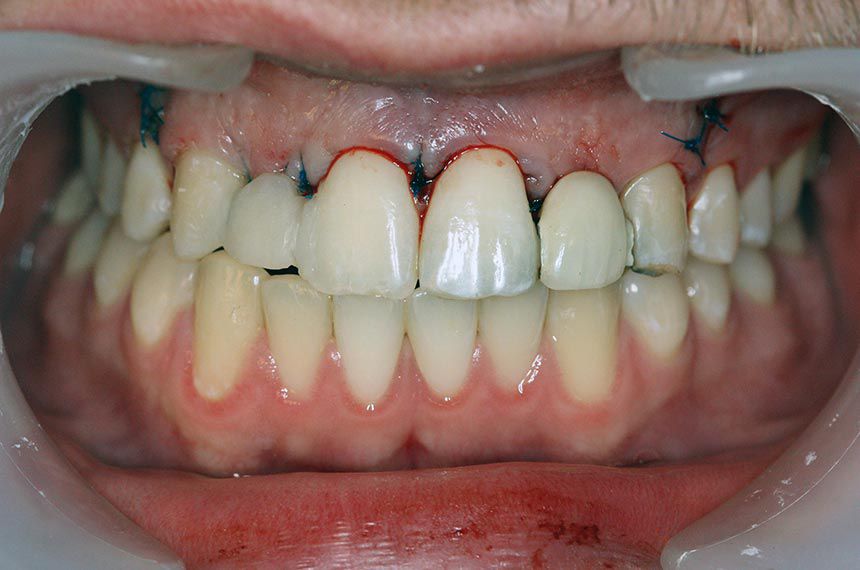
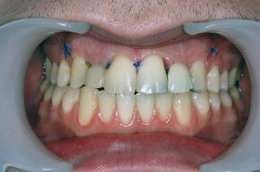
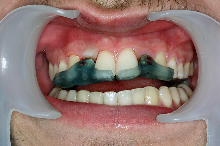
Surgical procedure

A marginal incision was made from the central incisor to the canine, including a releasing incision distally to avoid scar formation in the esthetic zone (Fig. 6). The mucoperiosteum was reflected (Figs. 7 – 8), and Straumann® BLT implants (Roxolid®, SLActive® 3.3mm/12mm) were placed in regions 12 (Figs. 9 – 11) and 22. The buccal bone was intact but needed horizontal augmentation to ensure long-term stability of the implant and to avoid grey shine through the gingiva and gingival recession. botiss cerabone® was added on the buccal side and covered with a botiss Jason® membrane. Emdogain® was added on top for better wound healing, and also because of its antibacterial activity (tooth position 12: (Figs. 12 – 14), tooth position 22: (Figs. 15 – 17). The periosteum was cut to obtain a tension-free primary closure (Figs. 18 – 19). Healing post-surgery was uneventful and, after 10 days, reached the status shown in (Figs. 20 – 21). The implants healed for three months before healing abutment surgery. The Maryland bridges were re-cemented in the meantime.

Prosthetic procedure

After three months a small incision was made on top of the implants, and the mucosa pushed to the buccal side. Provisional abutments were placed (Fig. 22), and composite was added to form the correct emergence profile. After a further two months of gingival healing, the final impression was taken and individual Straumann® CAD titanium abutments (for strength) were ordered, together with full zirconia crowns (for esthetic purposes) (Figs. 23 – 25). The crowns were cemented with temporary cement to make sure that the patient was fully satisfied with the esthetic result and, if this was the case, permanently cemented two weeks later.

Final result

The patient was very happy with the result (Figs. 26 – 29), which blended very naturally with the dentition as regards color and shape of both the crowns and the gingival portion. He only experienced slight swelling but no pain after implant surgery.